Restless legs syndrome (RLS) is a movement condition that causes a strong urge to move your legs when you’re resting. You may also feel sensations like itching, pulling, crawling or throbbing. There isn’t a cure for RLS, but treatment is available.
What is restless legs syndrome?
Restless legs syndrome (RLS) is a brain, nerve and sleep condition that causes a strong, nearly irresistible urge to move your legs that’s at least partially relieved by movement. Symptoms are more common when your body is at rest in the evening. It usually occurs in addition to uncomfortable sensations in your legs like throbbing or aching. The urge to constantly move can interfere with your ability to relax or fall asleep.
You may hear your healthcare provider call RLS restless leg syndrome or Willis-Ekbom disease.
What are the types of restless legs syndrome?
There are two types of RLS:
- Early onset: A diagnosis happens before age 45, it usually runs in your biological family history and the condition progresses slowly.
- Late onset: RLS progresses more quickly and a diagnosis happens after age 45.
How common is restless legs syndrome?
Between 7% and 10% of the United States population has restless legs syndrome. It’s more common among white people and females. Your risk of developing it increases as you get older.
What are the symptoms of restless legs syndrome?
Symptoms of restless legs syndrome include:
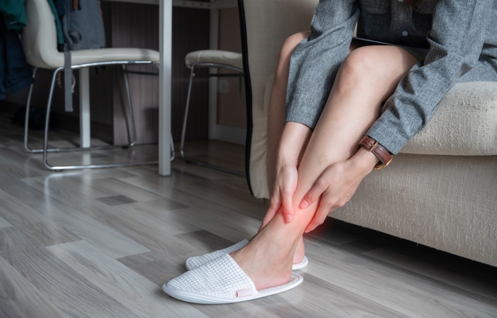
- Uncomfortable sensations in your legs that make you want to move them.
- Sensations get worse when you’re resting.
- Relief of discomfort (at least temporarily) when you move your legs.
- Twitchy legs or leg jerks in the evening and during sleep.
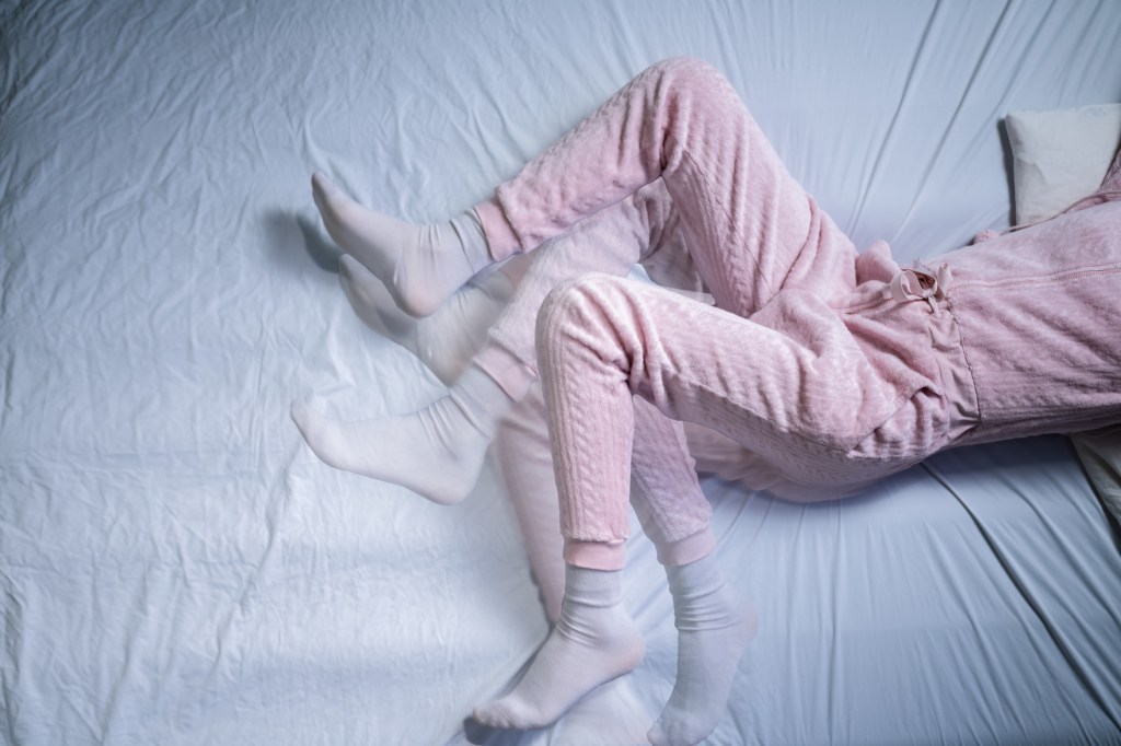
Symptoms of restless legs syndrome can affect your sleep. This can cause:
- Sleep disruptions, difficulty falling asleep or staying asleep.
- The urge to get out of bed to stretch or move your legs.
- Fatigue or daytime sleepiness.
- Behavior or mood changes.
- Difficulty paying attention, remembering things or concentrating.
- Depression or anxiety.
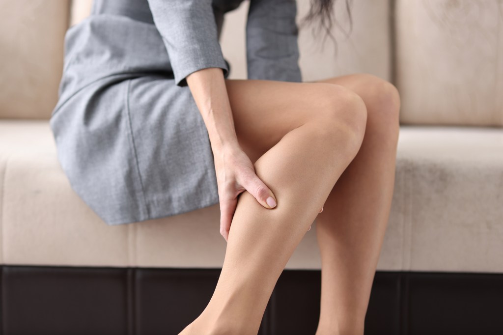
What does restless legs syndrome feel like?
Restless legs syndrome causes sensations that make you want to move your legs. These sensations most often happen in your legs, but they can also affect your arms or your entire body. You may experience the following feelings:
- Crawling.
- Itching.
- Aching.
- Burning.
- Throbbing.
- Pulling.
- Tugging.
Sensations can happen on one side of your body, but more commonly affect both sides equally, like both legs.
What causes restless legs syndrome?
The cause of restless legs syndrome isn’t well understood. It may relate to how the part of your brain that causes movement (basal ganglia) functions. The basal ganglia use dopamine to regulate how your body moves. If this part of your brain doesn’t get enough dopamine, it isn’t able to regulate your movement as efficiently as it should. This can lead to RLS symptoms.
Research suggests the following may contribute to RLS:
- Genetics: You can inherit RLS. During conception, one of your biological parents passes a genetic trait (autosomal dominant) that leads to an RLS diagnosis.
- Iron deficiency: Iron can be low in your brain despite normal blood levels.
- Underlying medical condition: Some conditions can cause secondary RLS, which is when RLS occurs with another medical condition.
- Medications: Certain medications, like antihistamines, antidepressants or antinausea medications, can cause RLS or make symptoms worse.
What conditions cause restless legs syndrome?
Some medical conditions can happen in addition to RLS, including:
- Low levels of iron (iron deficiency).
- Anemia.
- Pregnancy.
- Diabetes.
- Kidney disease.
- Peripheral neuropathy.
- Substance use disorder.
What triggers restless legs syndrome?
Triggers are things that make your RLS symptoms worse. Possible triggers can vary but could include:
- Alcohol.
- Caffeine.
- Nicotine.
- Certain medications.
- Stress.
If you experience these triggers or use/take them before you rest or go to bed, they’re more likely to set off your symptoms. In addition, a lack of sleep can lead to worsening symptoms. This means that your symptoms can trigger worsening symptoms. If you need help identifying what triggers your symptoms, talk to a healthcare provider.
What age group does restless legs syndrome affect?
Restless legs syndrome can affect anyone, including children, adolescents and adults. It’s more common to affect people after age 50. Symptoms tend to get worse as you age.
How is restless legs syndrome diagnosed?
A healthcare provider will diagnose RLS after a physical exam to review your symptoms. During the exam, they’ll take a complete medical history and family medical history.
As there isn’t a test to diagnose RLS, a healthcare provider may offer a neurological exam and blood tests to rule out other conditions or determine the cause of your symptoms. Your provider may recommend an overnight sleep study to evaluate other possible sleep conditions. However, RLS is a clinical diagnosis that doesn’t require sleep testing.
To confirm a diagnosis of RLS, a healthcare provider will look for the following criteria:
- You have the urge to move your legs, usually occurring with uncomfortable sensations like aching or pulling.
- Symptoms begin or worsen during periods of rest or inactivity.
- You have partial or total relief when stretching, walking or exercising the affected muscles.
- Your symptoms are worse or only occur in the evening or at night.
- Another medical condition didn’t cause your symptoms.
Is restless legs syndrome hard to diagnose in children?
Yes, it’s sometimes difficult for healthcare providers to diagnose RLS in children. This is because a child may not be able to describe their symptoms or what they feel. It’s common for RLS in children to look like attention-deficit/hyperactivity disorder (ADHD) or growing pains.
How is restless legs syndrome treated?
Treatment for RLS may include taking medications or changing your routine at home to help relieve your symptoms. Some people may reduce their symptoms if they work with their healthcare provider to manage other underlying health conditions. You and your healthcare provider will discuss the treatment options that might be best for you, as well as any side effects to look out for.
Restless legs syndrome medication
Certain medications can help relieve your symptoms of RLS. Your healthcare provider may recommend or prescribe the following:
- Iron supplements, taken with vitamin C.
- Antiseizure medications (gabapentin, pregabalin).
- Dopamine agonists (pramipexole, ropinirole).
- Dopamine precursors or medications that turn into dopamine (levodopa).
Certain medications like benzodiazepines (clonazepam), hypnotics (zolpidem) or opioids (methadone, buprenorphine) may help with severe cases if all other forms of treatment are ineffective. The side effects of these medications may be harmful and can lead to dependence on the medication.
There are many types of iron supplements. Blood tests can help to identify the best type for you. When levels of iron in your brain are suspected to be very low and RLS symptoms are severe, an iron infusion may help.
Medications that increase dopamine can worsen RLS symptoms over time (augmentation). Close monitoring is required.
At-home restless legs syndrome therapies
If you have mild RLS symptoms, a healthcare provider may recommend the following at-home therapies to help you feel more comfortable and fall asleep with restless legs. These may include:
- Getting regular exercise, such as aerobics, riding a bike/stationary bike or walking. Avoid heavy or intense exercise within a few hours of bedtime.
- Following good sleep habits like avoiding reading, watching television or being on a computer or phone while lying in bed. Not getting enough sleep can make RLS symptoms worse.
- Soaking your legs in a warm tub, and applying a heating pad or cold compress to your legs. These may provide temporary relief for your discomfort.
- Reducing your overall stress. A mental health professional can help you with this.
- Avoiding caffeine, like drinking coffee, before bedtime.
What relieves restless legs fast?
Moving your legs can temporarily relieve restless legs immediately, but your symptoms often return when you stop moving. You can also try massaging your legs, walking around or stretching.
Is restless legs syndrome serious?
Restless legs syndrome doesn’t affect your life expectancy, but it can affect your overall wellness. Symptoms may be mild to severe. Even mild symptoms can have a major impact on your life. Depending on what your definition of a “serious” medical condition is, your symptoms may fall into this category.
Is there a cure for restless legs syndrome?
There’s no cure available for RLS, but treatment can help manage your symptoms.
Can restless legs syndrome be prevented?
There’s no known way to prevent restless legs syndrome. You can reduce your risk of experiencing worsening symptoms by treating any underlying health conditions or avoiding triggers like caffeine and alcohol.
When should I see a healthcare provider?
Visit a healthcare provider if you have symptoms of RLS that:
- Don’t improve with at-home therapies.
- Get worse.
- Affect your ability to sleep.
What questions should I ask my healthcare provider?
- What caused my symptoms?
- What type of treatment do you recommend?
- Are there side effects of the treatment?
- How do I get better sleep with RLS?
- How can I improve my nighttime routine to decrease RLS symptoms?
A note from QBan Health Care Services
Living with restless legs syndrome (RLS) can be a constant annoyance. It interferes with your ability to fall asleep and stay asleep. When your alarm goes off in the morning, you may wish you could hit the snooze button a few more times. Drinking coffee (caffeine) to cope in the morning may make your symptoms worse at night. While there isn’t a cure available for RLS, treatment can help you manage your symptoms, feel better and get back to a regular sleeping routine.