
PERICARDITIS

Pericarditis is an inflammation of the pericardium or sac that contains your heart. Most people do well with prompt treatment, rest and medication. Others may need a procedure to drain fluid from their pericardium. In most cases, people make a full recovery. However, it can take weeks or months.

What is pericarditis?

Pericarditis is an inflammation of the pericardium, the thin, two-layered, fluid-filled sac that covers the outer surface of your heart. Pericarditis usually develops suddenly and may last from weeks up to several months. The condition usually clears up after three months, but sometimes attacks can come and go for years. Sometimes there is extra fluid in the space between the pericardial layers, which is called pericardial effusion.

Types of pericarditis

  • Acute pericarditis: Inflammation of the pericardium that develops suddenly along with the sudden onset of symptoms.
  • Chronic pericarditis: Inflammation of the pericardium that lasts for three months or longer after the initial acute attack.
  • Constrictive pericarditis: A severe form of pericarditis in which the inflamed layers of the pericardium stiffen, develop scar tissue, thicken and stick together. Constrictive pericarditis interferes with your heart’s normal function. This usually happens after multiple episodes of acute pericarditis over time.
  • Infectious pericarditis: develops as the result of a viral, bacterial, fungal or parasitic infection.
  • Idiopathic pericarditis: Pericarditis that doesn’t have a known cause.
  • Traumatic pericarditis: develops as the result of an injury to the chest, such as after a car accident.
  • Uremic pericarditis: develops as the result of kidney failure.
  • Malignant pericarditis: develops as result of a cancer growing in your body.

Myocarditis vs. pericarditis

Both of these are types of inflammation in your heart, but they’re in different places. Myocarditis is in your heart muscle. Pericarditis happens in your pericardium (the lining around your heart). Most often, a virus causes myocarditis and pericarditis. Both can give you chest pain, but with pericarditis, your chest pain should feel better when you sit up and lean forward. With myocarditis, you’ll usually feel tired and weak.

Who does pericarditis affect?

Pericarditis can affect anyone, but it’s most common in men and people assigned male at birth who are between the ages of 16 and 65. An estimated 28 people per 100,000 get pericarditis each year.

How does pericarditis affect my body?

When you have pericarditis, the membrane around your heart is red and swollen, like the skin around a cut that becomes inflamed. The pericardium is a thin, two-layered, fluid-filled sac that covers the outer surface of your heart. It provides lubrication for your heart, shields it from infection and malignancy, and contains your heart in your chest wall. It also keeps your heart from overexpanding when blood volume increases, which keeps your heart functioning efficiently.

What are the symptoms of pericarditis?

Pericarditis symptoms include chest pain that:

  • Is sharp and stabbing (This happens when your heart rubs against your pericardium).
  • May get worse when you cough, swallow, take deep breaths or lie flat.
  • Feels better when you sit up and lean forward.

You also may feel the need to bend over or hold your chest to breathe more comfortably.

Other pericarditis symptoms include:

  • Pain in your back, neck or left shoulder.
  • Trouble breathing when you lie down.
  • A dry cough.
  • Palpitations (feeling like your heart is racing or beating irregularly).
  • Anxiety or fatigue.
  • Fever.
  • Swelling of your legs, feet and ankles in severe cases.

Swelling in your feet, legs and ankles or shortness of breath every time you exert yourself may be a symptom of constrictive pericarditis. This is a severe type of pericarditis where the pericardium gets hard and/or thick. When this happens, the heart muscle can’t expand, which keeps your heart from working as it should. Your heart can become compressed, which makes blood back up into your lungs, abdomen and legs, leading to swelling and causing symptoms of congestive heart failure. You can also develop an abnormal heart rhythm.

If you have any symptoms of acute pericarditis, call your doctor right away. If you feel your symptoms are a medical emergency, call 911 right away to get treatment at the nearest hospital.

What causes pericarditis?

In up to 90% of cases, the cause of pericarditis is unknown. This is called idiopathic pericarditis.

There are many causes of pericarditis:

  • A complication of a viral infection, most often a gastrointestinal virus, causes viral pericarditis.
  • A bacterial infection, including tuberculosis, causes bacterial pericarditis.
  • A fungal infection causes fungal pericarditis.
  • An infection from a parasite causes parasitic pericarditis.
  • Some autoimmune diseases, such as lupus, rheumatoid arthritis and scleroderma, can cause pericarditis.
  • Injury to the chest, such as after a car accident, causes traumatic pericarditis.
  • Kidney failure causes uremic pericarditis.
  • Tumors like lymphoma causes malignant pericarditis.
  • Genetic diseases such as Familial Mediterranean Fever (FMF).
  • Medications that suppress the immune system (This is rare).

Your risk of pericarditis is higher after:

In these cases, it’s likely that the inflammation of the pericardium is an error in the body’s response to the procedure or condition. It can sometimes take several weeks for symptoms of pericarditis to develop after bypass surgery. Talk to your surgeon if you’re concerned about this.

How is pericarditis diagnosed?

Sharp pain in your chest and back of the shoulders that feel better when you sit up and lean forward, and chest pain with breathing are two major clues that you may have pericarditis and not a heart attack. Your healthcare provider will talk to you about your symptoms and medical history (such as whether you’ve recently been sick) and review your history of heart conditions, surgery and other health problems that could put you at a higher risk of pericarditis.

Your provider will listen to your heart. The rubbing of your pericardium’s inflamed lining causes a rubbing or creaking sound called the “pericardial rub.” They’ll be able to hear it best when you lean forward, hold your breath and breathe out. Depending on how bad the inflammation is, your provider may also hear crackles in your lungs, which are signs of fluid in the space around your lungs or extra fluid in your pericardium.

What tests will be done to diagnose pericarditis?

Healthcare providers use a variety of ways to check for pericarditis and any complications, such as pericardial effusion or constrictive pericarditis. You may need one or more tests, such as:

  • Chest X-ray to see the size of your heart and any fluid in your lungs.
  • Electrocardiogram (ECG or EKG) to look for changes in your heart rhythm. In about half of all people with pericarditis, providers see some characteristic changes to a normal heart rhythm. Some people don’t have any changes. If they do, they may be temporary.
  • Echocardiogram (echo) to see how well your heart is working and check for fluid (a pericardial effusion) around your heart. An echo will show the classic signs of constrictive pericarditis, including a stiff or thick pericardium that constricts your heart’s normal movement.
  • Cardiac MRI to check for extra fluid in your pericardium, pericardial inflammation or thickening, or compression of your heart. Your provider will give you a contrast agent called gadolinium during this highly specialized test.
  • CT scan to look for calcium in the pericardium, fluid, inflammation, tumors and disease of the areas around your heart. Your provider uses iodine dye during the test to get more information about the inflammation. This is an important test for patients who may need surgery for constrictive pericarditis.
  • Cardiac catheterization to get information about the filling pressures in your heart. This test can confirm a diagnosis of constrictive pericarditis.
  • Blood tests can help your provider make sure you’re not having a heart attack, see how well your heart is working, test the fluid in the pericardium and help find the cause of pericarditis. If you have pericarditis, it is common for your sedimentation rate (ESR) and ultra-sensitive C reactive protein levels (markers of inflammation) to be higher than normal. You may need other tests to check for autoimmune diseases like lupus and rheumatoid arthritis.

How is pericarditis treated?

Most times, people with pericarditis only need medications for pericarditis treatment, depending on the suspected cause. However, if you have a fluid buildup in your pericardium, you might need to have the fluid drained. If you have constrictive pericarditis, you may need surgery.

Medications for pericarditis

Treatment for acute pericarditis may include medication for pain and inflammation, such as ibuprofen or high-dose aspirin. Depending on the cause of your pericarditis, you may need an antibiotic or antifungal medication.

If you have severe symptoms that last longer than two weeks, or they clear up and then return, your healthcare provider may also prescribe an anti-inflammatory drug called colchicine (Colcrys® or Gloperba®). Colchicine can help control the inflammation and prevent pericarditis from returning weeks or even months later. Your provider may also prescribe a steroid medicine called prednisone, especially if you have kidney disease that makes it difficult for you to take ibuprofen and colchicine.

If you need to take large doses of ibuprofen, your provider may prescribe medications to ease gastrointestinal (stomach and digestive) symptoms. If you take large doses of nonsteroidal anti-inflammatory drugs (NSAIDs), you’ll need frequent follow-up appointments to look for changes in your kidney and liver function.

If you have chronic or recurrent pericarditis, you may need to take NSAIDs or colchicine for several years, even if you feel well. A diuretic (“water pill”) usually helps get rid of the extra fluid constrictive pericarditis causes. If you develop a heart rhythm problem, your provider will talk to you about treatment.

Your provider may also talk to you about treatment with steroids or other medications, such as azathioprine (Azasan® or Imuran®), IV human immunoglobulins or anakinra or rilonacept.

If your pericarditis is caused by an infection, your provider will prescribe specific medicines to treat that infection. If your pericarditis is caused by cancer, the most effective treatment is to treat the cancer.

Procedures and surgeries for pericarditis

When fluid builds up in the space between the pericardium, it can cause a condition called pericardial effusion. If the fluid builds up quickly, it can cause cardiac tamponade, a severe compression of the heart that impairs its ability to function. Cardiac tamponade is a medical emergency that requires prompt diagnosis and treatment.

This sudden buildup of fluid in between the layers of the pericardium keeps your heart from working like it should and can cause your blood pressure to drop. Because cardiac tamponade is life-threatening, your provider needs to drain the fluid immediately.

If fluid builds up in your pericardium (pericardial effusion) and compresses your heart, you may need a procedure called pericardiocentesis. Your provider uses a long, thin tube called a catheter to drain the extra fluid. Echocardiography or a CT scan helps guide the catheter and a needle to your pericardium.

If your provider can’t drain the fluid with a needle, they’ll perform a minimally invasive surgical procedure called a pericardial window. They’ll make an opening in the pericardium through a small chest incision to drain fluid from your pericardium.

If you have constrictive pericarditis, you may need to have some of your pericardium removed. This surgery is called a pericardiectomy. Surgeons perform this on people who develop scar tissue in their pericardium. It’s not normally for people who have active inflammation and chest pain from pericarditis.

Surgery isn’t usually used as a treatment for people with pericarditis that keeps coming back because inflammation makes healing after surgery difficult, but your provider may talk to you about it if other treatments aren’t successful.

How long does it take to recover from this treatment?

You should respond to treatment within a week, but you could be taking medicine for two weeks. Recovery from surgery takes longer. It can take weeks or months for a full recovery from pericarditis.

How can I reduce my risk?

Although you can’t prevent acute pericarditis, getting quick treatment and sticking with it can help you reduce your risk of getting it again. You should also follow your provider’s recommendations about when to start exercising again, as brisk exercise can worsen active pericarditis.

What can I expect if I have this condition?

You’ll need to take it easy while recovering from pericarditis. After you recover from pericarditis, you should be able to return to your normal activities without any reason for concern. Don’t return to vigorous exercise until your provider clears you. Your healthcare provider will talk to you about what to expect.

How long does pericarditis last?

Acute pericarditis lasts less than four to six weeks. Incessant pericarditis lasts longer than that but shorter than three months. Chronic pericarditis lasts more than three months. About 15% to 30% of people with pericarditis have repeat (or recurrent) episodes of pericarditis that come and go for many years.

Outlook for this condition

The outlook is very good for people with acute pericarditis who receive treatment. Most people make a full recovery. If you have a mild case, it may get better with rest. Without treatment, some people can end up with chronic pericarditis.

If bacteria or tuberculosis caused your pericarditis, you may have up to a 30% risk of constrictive pericarditis. Cardiac tamponade, a complication of pericarditis, is more likely to happen when cancer or infection causes your pericarditis.

How do I take care of myself?

It’s important to keep taking the medicines your healthcare provider prescribed and to keep all follow-up appointments you have with your provider.

When should I see my healthcare provider?

Contact your healthcare provider if you have symptoms of constrictive pericarditis, including:

When should I go to the ER?

Although chest pain is a common symptom of pericarditis, it’s also a symptom of a heart attack. Call 911 if you have chest pain because it could be a heart attack.

What questions should I ask my doctor?

  • Do you know what caused my pericarditis?
  • How long will I need to take medication?
  • What are the chances that I’ll get pericarditis again?

A note from QBan Health Care Services

If you get prompt treatment for pericarditis, you’ll most likely make a full recovery. Continuing with your treatment can help you prevent pericarditis from happening again. That’s why it’s important to keep taking prescribed medicines and go to all of your follow-up appointments. Get familiar with the symptoms of pericarditis so you can get quick treatment if it happens again.


CARDIOMYOPATHY

Cardiomyopathy refers to conditions that affect your heart muscle. If you have cardiomyopathy, your heart can’t efficiently pump blood to the rest of your body. As a result, you may experience fatigue, shortness of breath or heart palpitations. Cardiomyopathy gets worse over time. Treatment can slow the progression and improve your quality of life.

What is cardiomyopathy?

Cardiomyopathy is a disease that affects your myocardium (heart muscle). Cardiomyopathy can make your heart stiffen, enlarge or thicken and can cause scar tissue. As a result, your heart can’t pump blood effectively to the rest of your body.

In time, your heart can weaken and cardiomyopathy can lead to heart failure. Treatment can help. Some people with cardiomyopathy eventually need a heart transplant.

Types of cardiomyopathy

Types of cardiomyopathy include:

How common is cardiomyopathy?

Cardiomyopathy can affect anyone of any age, sex or race. The most common inherited cardiomyopathy, hypertrophic, affects about 1 in 500 people in the world. Other genetic types occur in 1 in 2,000 or 2,500 people.

What are the symptoms?

Cardiomyopathy symptoms include:

Some people have no symptoms of cardiomyopathy and don’t need treatment. Others experience symptoms as their disease progresses.

What causes cardiomyopathy?

Causes of cardiomyopathy include genes you get from your parents. Researchers have found thousands of genetic mutations that cause cardiomyopathies.

Other cardiomyopathy causes include:

Sometimes, experts don’t know the cause of a person’s cardiomyopathy.

What are the risk factors for cardiomyopathy?

Cardiomyopathy risk factors — some of which you can’t change — include:

What are the complications of cardiomyopathy?

As cardiomyopathy worsens, you may develop other heart problems, such as:

  • Arrhythmias (irregular heartbeats).
  • Heart failure.
  • Stroke.
  • Heart valve disease.
  • Cardiac arrest.
  • Cardiogenic shock.

How is cardiomyopathy diagnosed?

Your healthcare provider will ask about your medical history and that of your family. They’ll also do a physical exam.

If they suspect you have a heart condition, they’ll refer you to a cardiologist (heart specialist). A cardiologist performs a full assessment that may include a range of diagnostic tests.

What tests will be done to diagnose cardiomyopathy?

Tests to diagnose cardiomyopathy may include:

How is cardiomyopathy treated?

Cardiomyopathy treatments don’t cure the condition. However, they can help you manage your symptoms and slow down the disease’s progression. Your healthcare provider may recommend lifestyle changes, medications, devices or procedures. They’ll decide your treatment based on which type of cardiomyopathy you have and how advanced it is.

Lifestyle changes

Changing how you live day-to-day can strengthen your heart. Ways to do this include:

  • Eating food with low fat and salt.
  • Staying at a weight that’s healthy for you.
  • Getting regular exercise.
  • Getting the amount of sleep you need.
  • Reducing your stress level.
  • Avoiding the use of tobacco products.
  • Avoiding alcohol.

Medications

Depending on the cause, specific case, your cardiomyopathy and any other underlying medical conditions, certain medications can improve your blood flow and manage your symptoms.

Your cardiologist may prescribe:

Devices to correct arrhythmias

Devices a provider puts into your body can treat irregular heart rhythms. These devices monitor your heartbeat. They send electrical impulses to your heart when an arrhythmia starts. You may receive one of these devices:

Devices to improve blood flow

Some devices help your heart pump blood more efficiently. You may receive one of these devices:

Procedures

If you have severe symptoms or underlying heart conditions, your provider may recommend a procedure. Some of these involve surgery. Providers usually only recommend open-heart surgery or a heart transplant when all other treatments have failed to bring relief. Most people with cardiomyopathy don’t need the procedures below.

Rarely used procedures may include:

How can I lower my risk of cardiomyopathy?

You can take action to reduce your risk of conditions that could lead to cardiomyopathy.

Some of these actions include:

  • Managing your blood pressure.
  • Keeping your cholesterol within healthy ranges.
  • Getting regular exercise.
  • Avoiding tobacco products, alcohol and cocaine.
  • Managing underlying conditions such as sleep apnea or diabetes.
  • Scheduling regular checkups with a healthcare provider.
  • Taking all medications as prescribed.

You can’t prevent congenital (inherited) types of cardiomyopathy, but you can let relatives know that you have cardiomyopathy. Your relatives can talk to their providers to see if they’re at risk for it or have it. Early diagnosis helps people make a plan for managing cardiomyopathy.

What can I expect if I have cardiomyopathy?

Cardiomyopathy is a serious condition. Without treatment, it can be life-threatening. It’s also a progressive condition, meaning it usually worsens over time.

Cardiomyopathy treatment can improve your outlook. If you have cardiomyopathy, seek treatment from a cardiologist (heart specialist). Medication, procedures or other treatments can increase your quality of life and help you live longer.

How long cardiomyopathy lasts

The length of time that you have cardiomyopathy depends on the type you have. The severity of your condition matters, too. Takotsubo cardiomyopathy is usually temporary and doesn’t last long. Cardiomyopathy from alcohol may only last a few months. You may be able to reverse cardiomyopathy from chemotherapy.

For other types, you’ll need to manage your symptoms for the rest of your life. Many people don’t have major problems from their cardiomyopathy, but some do.

Outlook for cardiomyopathy

There’s no cure for cardiomyopathy. However, you can manage the condition or slow its progression. Many people who make healthy lifestyle choices and seek medical treatment can live a high quality of life with cardiomyopathy. Taking the proper medications can drastically alter and improve the prognosis of cardiomyopathy. Many people can live normal and healthy lives with the right treatment.

How do I take care of myself?

Lifestyle choices can affect how quickly cardiomyopathy progresses. To manage symptoms and increase your heart health, your provider may recommend:

When should I see my healthcare provider?

You’ll need regular checkups to monitor your heart’s health. Also, you may need regular blood tests or other tests that tell your provider how well your heart is doing. Contact your provider if you have:

  • New symptoms.
  • Worse symptoms.
  • Side effects from medications.
  • Trouble with your device.

When should I go to the ER?

Get emergency medical care if you have:

  • Chest pain.
  • Heart palpitations.
  • Fainting.

What questions should I ask my doctor?

You may want to ask your healthcare provider:

  • What’s the most likely cause of my cardiomyopathy?
  • What treatments do you recommend?
  • What lifestyle changes should I make to improve my heart health?
  • Is there a support group nearby or online that I can join?

A note from QBan Health Care Services

It’s normal to have concerns about a cardiomyopathy diagnosis. But healthcare providers can offer many treatments to improve your quality of life and help you live longer. Ask your provider how often you need checkups with them. Going to regular appointments with your provider will help them monitor your condition and see how well treatments are working for you.

MEDICAL SCANS

Have you ever had to get an X-ray, MRI, or other medical scan? Do you know what these tests involve? Or what they can do?

Medical scans help doctors diagnose everything from head trauma to foot pain. There are many different types of imaging technologies. Each works differently.

Some types of imaging tests use radiation. Others use sound waves, radio waves, or magnets. Learning about how medical scans work can help you feel more comfortable if you or a loved one needs one. It can also help you to know what to ask about before getting an imaging test.

Radiation in Medical Scans

Radiation can be dangerous, but it can also save lives. How can that be? Harnessed properly, radiation can help diagnose and even treat disease. So when you’re faced with a medical test that uses radiation, don’t let fear get in your way. Learn about the risks and benefits, and know what questions to ask. If medical radiation is really needed, take steps to ensure that it’s done as safely as possible.

Radiation, simply put, is the transfer of energy through space. The energy may be in the form of invisible particles or waves. Radiation is all around us—and has been throughout our evolution—so our bodies are designed to deal with the low levels we’re exposed to every day. Excess radiation, however, can damage tissues and lead to serious problems.

Why is radiation used in medicine? Radiation allows radiologists and other physicians to see internal parts of the body that they aren’t able to see directly with their eyes or through other physical examination measures.

Techniques like x-rays and CT scans send controlled amounts of radiation through the body and create images based on what comes through the other side. Another imaging method called nuclear medicine uses compounds that emit radiation, which can then be detected outside the body. Injected or swallowed, these compounds can target a specific area and reveal internal problems. Or they can be used to track how well internal organs are working.

With these advanced imaging tools, doctors can detect disease early, when it’s easier to treat. As a result, use of medical radiation has been rising. But with these benefits come some risks.

One of the main risks of being exposed to radiation is the possibility of developing a cancer. Cancer takes years to develop, so it’s nearly impossible to tell exactly what causes any given cancer. As a result, it’s hard for researchers to gauge exactly how much risk a given amount of radiation poses.

Children’s growing bodies are even more susceptible to radiation damage than those of adults. Children have a long lifetime ahead. Any damage caused by radiation has a longer period of time to show itself.

In 2012, a worrisome report linked childhood CT scans to 2 types of cancer later in life: leukemia and brain cancer. The NIH-funded study looked at more than 175,000 children and young adults. Depending on the radiation dose, those who’d received 2 or more scans had a small boost in cancer risk. Because these cancers are rare, the benefits of CT scans likely outweigh the risks, the researchers concluded. Still, they suggest that doctors consider alternatives to CT scans or use the lowest possible radiation doses.

Strangely enough, radiation can also be used to treat cancer. Focused doses of high-energy radiation can kill cancer cells and shrink tumors. Medical imaging tests, in contrast, use much lower doses.

The fundamental thing to understand is that these procedures all deliver a generally low amount of dose. In some cases, depending on your disease, it’s potentially riskier to not get the scan or nuclear medicine procedure than to get it.

If a doctor recommends a test that uses radiation, ask about its risks and benefits. If the test is truly needed, do some research into the imaging facilities. Find one that monitors the doses they’re giving patients and takes pride in low doses.

X-Rays

The first revolution in seeing into the body came with X-rays. They have been used in the clinic for more than 120 years.

X-rays are still used every day because they can do a lot. They’re useful for looking at bones and finding problems in certain types of tissues, like pneumonia in the lungs.

X-ray imaging works by passing an energy beam through a part of your body. Your bones or other body parts will block some of the X-ray beams from passing through. That makes their shapes appear on the detectors used to capture the beams. The detector turns the X-rays into a digital image for a radiologist to look at.

X-ray beams use radiation. Radiation is energy that’s released as invisible particles or waves. Being exposed to very large amounts of radiation can damage cells and tissues. It may also increase your risk of developing cancer.

But modern X-ray tests use a very small dose of radiation. People are naturally exposed to radiation from many sources, such as the sky, rocks, and soil.

A chest X-ray gives you similar amounts of radiation as you’d get in a plane flight across the Atlantic Ocean.

CT Scans

CT scans also use X-ray beams. But the beams rotate around your entire body to create a 3D picture. These images contain more information than a regular X-ray. The scan can be done in less than a minute. That makes it especially useful in places like the emergency department. There, doctors need to know immediately if a patient has a life-threatening condition.

Because CT scans use more X-ray beams than a normal X-ray, they often deliver a higher dose of radiation. But medical specialists have ways to calculate the smallest radiation dose needed.

The medical specialists tailor the dose to the patient’s size, and they tailor it to the reason for the exam. For example, a CT scan of the chest needs less radiation than a CT scan of the stomach area.

While lower doses of radiation would likely further lower risk, the standard doses are already quite low. That’s important for people to know, because some patients who really need a CT scan are afraid to get it.

Fear can sometimes keep someone from getting a scan that could help improve their health, or even save their life. Current CT doses are in a range where it’s not possible to even prove a risk exists. They’re that low.

MRI

MRI works in a very different way. It doesn’t use X-rays. Instead, it uses strong magnets and radio waves to affect atoms in the water molecules within your body’s tissues. When the radio waves are turned off, the atoms release energy that’s detected by the MRI machine.

Atoms in different tissue types go back to normal at different speeds and release different amounts of energy. MRI software uses this information to create 3D pictures of the different tissue types.

MRI is most helpful when you want to look at diseases that involve soft tissue, such as muscles, tendons, and blood vessels.

MRI can provide information about how the body is functioning in real time. For example, we can measure how much blood is flowing in the vessels. That can help doctors find small blockages or defects in the heart.

Because MRI doesn’t use X-rays, doctors would like to use it more in children. But MRI machines require you to lie motionless for a long time.

It can be difficult for children to hold still. If needed, general anesthesia can help get kids through the test. It makes them unconscious and unable to move. It’s typically very safe, but comes with some risks.

To help reduce the use of anesthesia, medical specialists have created a flexible, blanket-like version of MRI hardware to use with children. They coupled it with new methods for faster scanning. The soft blanket-like coil sits closely on top of the patient, providing a comforting environment. “It’s helping some kids get though exams without anesthesia.

Ultrasound

Another commonly used imaging method is called ultrasound. It is a noninvasive test that sends sound waves into the body. Different types of tissue reflect sound waves differently. These differences can be picked up by an ultrasound machine and turned into a picture or video. Ultrasound is helpful for looking at the heart and other internal organs, other soft tissues such as blood vessels, or a developing baby.

Ultrasound enables healthcare providers to “see” details of soft tissues inside your body without making any incisions (cuts). And unlike X-rays, ultrasound doesn’t use radiation.

Although most people associate ultrasound with pregnancy, healthcare providers use ultrasound for many different situations and to look at several different parts of the inside of your body.

Other Scans

Doctors also use tests called nuclear imaging. These tests use a tiny amount of a radioactive substance, or “tracer.” Most tracers are injected into the body, but some are inhaled or swallowed. The tracers inside the body release radiation that can be measured by a detector outside the body. The type of tracer differs depending on what the doctors want to see.

A positron emission tomography (PET) scan, for example, often uses a radioactive sugar to diagnose cancer. When cancer cells take up the radioactive sugar, they can be seen with the PET scanner.

Scientists are working to develop new types of tracers to detect different conditions, such as infections hiding deep in the body. They’re also continuing to explore other ways to make medical scans faster and deliver less radiation.

A Note from QBan Health Care Services

Medical scans are very useful and generally safe imaging tests that healthcare providers use for a variety of reasons. If you need a medical scan and are worried about the exam or have questions about it, don’t be afraid to ask your healthcare provider. Talk to them about any concerns you have, including all options for testing and the benefits and risks of each. They’re available to help and support you.

CONGENITAL HEART DISEASE

Congenital heart disease (CHD) is a defect in the heart’s structure that’s present at birth. It can be detected before birth, soon after birth or anytime throughout life. There are many kinds of CHD. Symptoms and treatment depend on the type of defect and its severity. A person with CHD should see a cardiologist regularly throughout life.

What is congenital heart disease?

Congenital heart disease (CHD) is a defect or problem with the heart’s structure that’s present at birth, such as:

  • A hole in the heart wall.
  • Issues with the blood vessels (too many or too few, blood flowing too slowly, to the wrong place or in the wrong direction).
  • Problems with the heart valves.

Some cases of CHD are simple and may not cause any symptoms, but others can be life-threatening and require treatment.

Heart defects may be detected early (before a baby is born or shortly afterward). But sometimes, CHD isn’t diagnosed until childhood, adolescence or adulthood.

How common are congenital heart defects?

CHD is the most common type of birth defect, affecting 8 to 9 per 1,000 live births.

What are the types of congenital heart defects?

There are two main groups of CHD: cyanotic congenital heart disease and acyanotic congenital heart disease.

Cyanotic (low oxygen in the blood) congenital heart disease

Cyanotic congenital heart disease involves heart defects that reduce the amount of oxygen delivered to the rest of the body. This is sometimes called a critical congenital heart defect. Babies born with cyanotic congenital heart disease usually have low levels of oxygen and need surgery. Examples include:

  • Left heart obstructive lesions: This type of defect reduces blood flow between the heart and the rest of the body (systemic blood flow). Examples include hypoplastic left heart syndrome (when the heart is too small on the left side) and interrupted aortic arch (aorta is incomplete).
  • Right heart obstructive lesions: This type of defect reduces blood flow between the heart and lungs (pulmonary blood flow). Examples include tetralogy of Fallot (a group of four defects), pulmonary atresia and tricuspid atresia (valves don’t develop correctly).
  • Mixing lesions: With this type of defect, the body mixes systemic and pulmonary blood flow. One example is transposition of the great arteries when the two main arteries leaving the heart are reversed. Another is truncus arteriosus, when the heart has only one main artery, instead of two, to carry blood to the body.

Acyanotic (blood oxygen level is acceptable) congenital heart disease

Acyanotic congenital heart disease involves a defect that causes blood to pump through the body abnormally. For example:

  • Hole in the heart: One of the heart’s walls can have an abnormal opening. Depending on the location of the hole, this may be called atrial septal defect, atrioventricular canal, patent ductus arteriosus or ventricular septal defect.
  • Problem with the aorta: The aorta is the main artery that carries blood away from your heart to the rest of the body. It can be too narrow (aortic coarctation). Or the aortic valve (which opens and closes to regulate blood flow) may be restricted in opening and or a more common defect with only two flaps instead of three (called bicuspid aortic valve).
  • Problem with the pulmonary artery: The pulmonary artery carries blood from the right side of the heart to the lungs to get oxygen. If this artery is too narrow, it’s called pulmonary artery stenosis.

What are the symptoms of congenital heart disease?

Symptoms may start as soon as a baby is born or may not appear until later in life. They can include:

  • Cyanosis (blueish skin, lips or nails).
  • Excessive sleepiness.
  • Fast breathing or trouble breathing.
  • Fatigue (extreme tiredness).
  • Getting unusually tired or out of breath during exercise.
  • Heart murmur (a swishing sound made by the heart that may indicate abnormal blood flow).
  • Poor blood circulation.
  • Weak pulse or pounding heartbeat.

The signs and symptoms vary widely, depending on:

  • Age.
  • The number of heart defects (a person can be born with more than one defect).
  • The severity of the condition.
  • Type of defect.

What causes congenital heart disease?

CHD happens when the fetal heart doesn’t develop correctly in the uterus. Scientists don’t fully understand why that happens, but it may be related to:

  • Abnormal chromosomes or genetics.
  • Drinking or smoking during pregnancy (or significant environmental exposures such as secondhand smoke).
  • Illnesses in the mother during pregnancy (diabetes, drug use, phenylketonuria or viral infection).

How is congenital heart disease diagnosed?

Sometimes a heart defect is found before a baby is born. If your healthcare provider finds anything unusual during a routine prenatal ultrasound, you and the fetus may need further testing. For example, a fetal echocardiogram uses sound waves to create pictures of the fetal heart.

Other heart defects are found soon after a baby is born. For example, cyanotic CHD is often detected by pulse oximetry. The simple, painless test uses sensors on the fingers or toes to find out if oxygen levels are too low. Sometimes, congenital heart disease isn’t diagnosed until later in life.

Tests that can help diagnose CHD in newborns, children or adults include:

  • Physical exam: During an exam, a healthcare provider will listen to the heart for any abnormal sounds.
  • Chest X-ray: A chest X-ray takes pictures of the inside of your chest to reveal any structural abnormalities.
  • Electrocardiogram: Electrocardiogram (EKG or ECG) measures the electrical activity of your heart.
  • Echocardiogram: An echocardiogram (echo) uses ultrasound to create images of the heart’s valves and chambers.
  • Heart catheterization: A healthcare provider can tell how well your heart is pumping and circulating blood by performing a heart catheterization. It’s also called cardiac catheterization or coronary angiography.
  • Magnetic resonance imaging (MRI): MRI for heart disease can create detailed pictures of your heart.

How is CHD treated?

Some cases of CHD may heal themselves. Others may remain but don’t require any treatment while others must be treated soon after birth.

Anyone with a heart defect should see a heart specialist (cardiologist) regularly throughout their life. Be sure the cardiologist has training and expertise in congenital heart defects.

Treatment for CHD may involve:

  • Catheter procedure to place a plug into the defect.
  • Medications to help your heart work more efficiently or to manage blood pressure.
  • Nonsurgical procedure to close a defect using a closing device.
  • Oxygen therapy, which provides higher levels of oxygen than normal room air would.
  • Prostaglandin E1, which relaxes smooth heart muscle and can keep open the ductus arteriosus (a blood vessel normally closed after birth) helping provide needed circulation.
  • Surgery to repair a defect, open up blood flow or redirect blood. In severe cases, a heart transplant may be necessary.

Does adult congenital heart disease (ACHD) require special treatment?

Adults with congenital heart disease may develop certain health problems later in life. They can limit your ability to perform everyday tasks and shorten your life span. Examples include:

  • Arrhythmia: Arrhythmia is an abnormal heart rhythm. It can occur when there is a problem with the heart’s structure or scar tissue from an earlier surgery.
  • Endocarditis: Endocarditis is an infection in the heart. Taking antibiotics if indicated for a specific defect before dental and surgical procedures can help prevent heart infection.
  • Heart failure: In heart failure, the heart can no longer pump adequately for proper blood circulation to fulfill the body’s needs. It’s sometimes called congestive heart failure.
  • Hypertension: Hypertension (high blood pressure) occurs when pumping blood puts too much pressure on blood vessel walls.
  • Pregnancy complications: Women with ACHD may have an increased risk of pregnancy complications, including arrhythmia, heart failure and stroke.
  • Pulmonary hypertension: Pulmonary hypertension is high blood pressure in the lung’s arteries. It can lead to heart failure.
  • Stroke: A stroke occurs when a blood vessel in the brain becomes blocked or bursts. This blockage or rupture cuts off blood supply to the brain.
  • Sudden cardiac death: Sudden cardiac death is a sudden loss of heart function. It’s caused by sudden cardiac arrest. This life-threatening condition occurs when the heart’s electrical system malfunctions, most commonly causing a dangerously fast heartbeat.
  • Valve dysfunction: If the heart’s valves aren’t working correctly, blood may be restricted or back up into the heart chambers causing overload or the heart to work harder than it should.

It’s important to see a cardiologist regularly to monitor your heart and prevent or treat complications. Treatment will depend on the type of heart defect you have and how it’s affecting you personally.

How can I reduce the risk of a heart defect?

Scientists don’t have all the answers yet as to what causes heart defects other than random gene mutations. But, there are things that place you at higher risk (smoking, alcohol, certain medications, etc.) and these should be avoided during pregnancy. There are otherwise no proven strategies to prevent CHD.

You should follow your healthcare provider’s instructions during pregnancy, including:

  • Don’t use recreational drugs.
  • Get all recommended screening tests during pregnancy to detect problems as early as possible.
  • Manage any health conditions, such as diabetes and phenylketonuria.
  • Stop smoking and avoid secondhand smoke.
  • Avoid alcohol.

What is the outlook?

The outlook for people with congenital heart disease depends on the type of defect and its severity. Although serious cases can be life-threatening, many people with CHD live long, relatively normal and fulfilling lives. Decades ago, only 10% of children with CHD survived into adulthood. Advances in diagnosis and treatment now help about 90% survive.

How do I take care of myself with adult congenital heart disease?

To keep your heart as healthy as possible and prevent complications of ACHD:

  • Eat a well-balanced, nutritious diet of heart-healthy foods.
  • Exercise regularly (but only with your cardiologist’s approval).
  • Maintain a healthy weight.
  • See a cardiologist regularly throughout your life to monitor and manage ACHD and detect any complications.
  • Tell all of your healthcare providers about the heart defect, all medications you take and what surgeries you’ve had.
  • Understand the specific type of heart defect you have and what the possible complications are.
  • If you plan to become pregnant, talk to your cardiologist, obstetrician and primary care doctor well in advance. They will help you understand and manage the risks throughout your pregnancy.

A note from QBan Health Care Services

Congenital heart disease is a defect in the heart’s structure that’s present at birth. It can be detected before a baby is born, soon after birth, or any time during childhood or adulthood. If you or your baby have a heart defect, it’s important to see a cardiologist who specializes in CHD. You should visit the cardiologist regularly throughout life to monitor the condition and keep the heart as healthy as possible.

EDEMA

Edema occurs when fluid builds up in your tissues, often in your feet, legs and ankles. Edema can affect anyone, especially people who are pregnant and adults age 65 and older. Treatment involves lifestyle changes, including diet and exercise.

What is edema?

Edema is the medical term for swelling caused by fluid trapped in your body’s tissues. Edema happens most often in your feet, ankles and legs, but can affect other parts of your body, such as your face, hands and abdomen.

Who does edema affect?

Edema can affect anyone, but the condition most often affects people who are pregnant and adults who are 65 years or older.

How common is edema?

Edema is common because there are many causes associated with the condition. Mild cases of edema go away on their own, so the exact rate of occurrence is unknown.

How does edema affect my body?

Edema will cause parts of your body to increase in size (swell), which might prevent you from completing your daily tasks. Simple lifestyle changes like elevating the swollen part of your body or moving around if you were sitting or standing for a long period of time can reduce swelling and help you feel better. Sometimes edema is a symptom of an underlying health condition, so contact your healthcare provider if you experience symptoms of edema.

What are the symptoms of edema?

A symptom of edema is swelling in your body. Swelling occurs when a part of your body gets bigger because there is a buildup of fluid in your tissues. Swelling can happen anywhere on your body but most often affects your feet, ankles and legs.

Symptoms of swelling include:

  • An area of your body is larger than it was a day ago.
  • The skin over the swollen area looks stretched and shiny.
  • Difficulty walking if your legs, ankles or feet swell.
  • You may be coughing or have trouble breathing.
  • You feel full or tightness in your swollen body part.
  • Mild pain or a sore feeling in the affected area.

What causes edema?

After your healthcare provider makes an edema diagnosis, their next step is to identify what caused fluid to build up in your tissues. There are several possible causes for an edema diagnosis including:

  • Gravity: If you spend a lot of time sitting or standing in one place for too long, water naturally pulls down into your arms, legs and feet (dependent edema).
  • Weakened valves of your veins (venous insufficiency): When the valves in your veins are weak, it is hard for your veins to push blood back up to your heart, and leads to varicose veins and a buildup of fluid in the legs.
  • Underlying medical conditions: Conditions like heart failure and lung, liver, kidney and thyroid diseases have edema as a symptom.
  • Side effects from medication: Some drugs, like blood pressure or pain management medications, have edema as a side effect.
  • Poor nutrition: If you aren’t eating a well-balanced diet or if you eat a lot of foods high in salt (sodium), fluid could build up in different parts of your body.
  • Pregnancy: Swelling in your legs during pregnancy occurs as the uterus puts pressure on your blood vessels in the lower trunk of your body.
  • Compromised immune system: An allergic reaction, infection, burns, trauma or clots can lead to edema.

How is edema diagnosed?

Your healthcare provider will give a physical examination to diagnose edema, followed by diagnostic tests to find the cause. They will look for swelling, especially on parts of your body where your skin has a shiny or stretched appearance.

What is edema grading?

Edema grading is a scale used to identify the severity of your edema diagnosis and estimate how much fluid built up in your tissues.

Your healthcare provider will test an area of your body for edema by gently pressing their finger on a swollen area of your skin for five to 15 seconds (pitting test). After they release pressure, a dimple (pit) will appear in your skin. The pit indicates that there is fluid built up in your tissues.

The edema grading scale measures how quickly the dimple goes back to normal (rebound) after a pitting test. The scale includes:

  • Grade 1: Immediate rebound with 2 millimeter (mm) pit.
  • Grade 2: Less than 15-second rebound with 3 to 4 mm pit.
  • Grade 3: Rebound greater than 15 seconds but less than 60 seconds with 5 to 6 mm pit.
  • Grade 4: Rebound between 2 to 3 minutes with an 8 mm pit.

How is edema treated?

Treatment for edema varies based on the cause, especially if the cause relates to an underlying health condition. For example:

  • If lung disease causes edema, such as emphysema or chronic bronchitis, your healthcare provider will recommend quitting smoking if you smoke.
  • If edema occurs with chronic heart failure, your provider will recommend lifestyle changes to treat your diagnosis by monitoring your weight, fluid intake and salt intake. Your provider might recommend cutting back on the amount of alcohol you drink.
  • If edema is a side effect of a medication you are taking, your provider might stop or lower the dosage of your medication to resolve the swelling. Do not stop taking your medication unless your provider tells you to do so.

Treatment to reduce swelling

In addition to treating the underlying cause of edema, there are a few steps you can take to keep fluid from building up in your body:

  • When you are sitting or lying down, put a pillow under your legs to keep them elevated above the level of your heart.
  • Do not sit or stand for long periods without moving or go on short walks.
  • Wear support socks, stockings or sleeves, which put pressure on parts of your body to keep fluids from collecting there. Edema shoes are available for people who experience chronic edema and need adjustable footwear for swelling.
  • Reduce the amount of salt in your diet.
  • Follow your doctor’s directions for taking medications. Your doctor might want you to take a diuretic (commonly called a “water pill”), which helps your body get rid of excess fluid.

What can’t I eat with edema?

In some cases, the cause of edema could be too much salt in your diet. Salt causes your body to retain water, which could leak into your tissues and cause swelling. Making lifestyle changes to reduce the amount of salt in your diet could improve your edema diagnosis.

How soon after treatment will I feel better?

Depending on the cause of your diagnosis, edema could be temporary or permanent. Swelling normally lasts for a few days. In the first two days, you will experience the most swelling, and it should start to reduce by the third day. Following treatment from your healthcare provider reduces the amount of swelling you might experience. If your swelling doesn’t go away after a few days of treatment, talk to your healthcare provider.

How can I prevent edema?

Sometimes, you can’t prevent what caused edema if it is the result of an underlying health condition like heart failure, liver or kidney disease, but you can work with your healthcare provider to manage symptoms.

If the cause of edema is too much salt intake, adjusting your diet to reduce the amount of salt in the foods you eat will prevent edema.

You can also prevent edema by moving around more frequently. Sitting or standing without moving could cause fluid to build up in your tissues. If you notice you’ve been sitting for a long period of time and you’re able to, get up or move your body around; it will reduce the likelihood of swelling.

What can I expect if I have edema?

It’s very important to see your healthcare provider if you experience edema or swelling in your body. Edema can stretch your skin and if not treated, swelling could increase and cause serious health problems.

Edema can be a short-term or long-term condition, depending on its cause. Treatment is available to help you manage any underlying conditions that might cause edema or you can make simple lifestyle changes to reduce swelling and fluid buildup in your body.

How do I take care of myself?

If you have edema, take steps to reduce swelling by:

  • Making lifestyle changes to stop smoking or changing your diet.
  • Moving around more often.
  • Elevating your legs when lying down or sitting.
  • Wearing compression socks, sleeves or stockings.

It is important to protect any swollen areas of your body from additional pressure, injury and extreme temperatures. Injury to the skin over swollen areas takes longer to heal and is more likely to become infected.

When should I see my healthcare provider?

Call your healthcare provider immediately if you experience:

  • Pain or discolored skin in a swollen area.
  • An open sore on a swollen area.
  • Shortness of breath.
  • Swelling of only one limb.
  • Difficulty walking or you have trouble moving.

What questions should I ask my doctor?

  • What caused my edema?
  • Do I need to reduce the amount of salt in my diet?
  • Are there side effects to the treatment?
  • Do I need to wear compression socks to reduce swelling in my ankles?

A note from QBan Health Care Services

Edema is common and ranges in severity for each person diagnosed with the condition based on the cause. If you are pregnant, it is normal to experience swelling as your due date nears. Normally, edema will go away on its own if you have a mild case, and medication and treatment are available if you have a more severe case.

If are not pregnant and you notice that you have unexpected swelling in a part of your body, contact your healthcare provider for an exam. Edema could be a sign of an underlying health condition and early diagnosis and treatment could lead to the best prognosis.

GOUT

Gout is a painful form of arthritis. When your body has extra uric acid, sharp crystals can form in your joints (usually your big toe). Flare-ups of symptoms like pain and swelling come and go in periods called gout attacks. Treatment is usually a combination of symptom management and changing your diet.

What is gout?

Gout is a form of inflammatory arthritis that causes pain and swelling in your joints. Gout happens when there’s a buildup of uric acid in your body.

Gout most commonly affects your big toe joint. But it can affect other joints, including your:

Gout symptoms come and go (recur) in episodes called flares or gout attacks. A healthcare provider will suggest medications and changes to your diet that will lower your uric acid levels and minimize how often you experience gout attacks in the future.

What are gout symptoms?

Gout attacks are very painful and can happen suddenly, often overnight. During a gout attack, symptoms in your affected joints may include:

  • Intense pain.
  • Discoloration or redness.
  • Stiffness.
  • Swelling.
  • Tenderness, even to a light touch (like a bedsheet covering your affected joint).
  • Warmth, or a feeling like the joint is “on fire.”

How long does a gout attack last?

Gout attacks usually last a week or two. You might have some flares that last longer than others, and some might cause more severe symptoms. Between attacks, you might not experience any gout symptoms.

What causes gout?

A buildup of excess uric acid in your body causes gout. Your body naturally makes uric acid when it breaks down chemicals called purines found in certain foods and drinks. Your kidneys usually filter uric acid out of your blood, and then it leaves your body when you pee. Sometimes your body makes too much uric acid, or your kidneys don’t remove it from your blood fast enough. When your body has high levels of uric acid (hyperuricemia), uric acid crystals can build up and settle into your joints. The sharp crystals clump together and cause sudden episodes of pain, swelling and other symptoms. Having temporarily high uric acid levels doesn’t mean you’ll definitely develop gout. Many people with hyperuricemia never get gout.

Gout risk factors

Gout can affect anyone. People assigned male at birth (AMAB) are three times more likely to develop gout. People assigned female at birth (AFAB) usually don’t experience gout until after menopause. People with certain health conditions are more likely to develop gout, including:

You’re more likely to experience gout if you:

  • Have a biological parent or grandparent who has gout.
  • Eat a lot of animal proteins — especially animal flesh, shellfish and foods that contain organ meat.
  • Drink alcohol regularly.
  • Take a diuretic medication (water pills).
  • Take immunosuppressants.

Which foods cause gout?

Eating or drinking foods high in purines are more likely to lead to high uric acid levels in your body that cause gout, including:

  • Sugary drinks and sweets: Standard table sugar is half fructose (fruit sugar), which breaks down into uric acid. Any food or drink with high sugar content can trigger gout.
  • High fructose corn syrup: This is a concentrated form of fructose. Packaged food products and processed snacks can contain lots of high fructose corn syrup.
  • Alcohol: Even though not all alcoholic drinks are high in purines, alcohol prevents your kidneys from eliminating uric acid, pulling it back into your body, where it continues to accumulate.
  • Organ meats: These include liver, tripe, sweetbreads, brains and kidneys.
  • Game meats: Specialties such as goose, veal and venison all contain high levels of purines.
  • Certain seafood: Herring, scallops, mussels, codfish, tuna, trout and haddock.
  • Red meat: Beef, lamb, pork and bacon.
  • Turkey: Especially processed deli turkey.
  • Gravy and meat sauces.

How is gout diagnosed?

A healthcare provider will diagnose gout with a physical exam. They’ll ask you about your symptoms and examine your affected joints. Tell your provider when you first noticed symptoms like pain and swelling in your joint and how often the symptoms come and go.

What tests will be done to diagnose gout?

Your healthcare provider might use a few imaging tests to take pictures of your affected joints. These tests can also show if gout has caused any changes in your joints. You might need:

Other common tests to diagnose gout include:

  • Blood tests to measure the uric acid in your blood.
  • Joint aspiration — using a needle to remove a sample of fluid from inside a joint.

How is gout treated?

Treating gout is usually a combination of managing your symptoms during a flare and reducing how often you consume high-purine foods and drinks.

Gout medication

Your healthcare provider might suggest medications to help manage your symptoms, including:

  • NSAIDs: Over-the-counter (OTC) NSAIDs, like ibuprofen and naproxen, can reduce pain and swelling during a gout attack. Some people with kidney disease, stomach ulcers and other health problems shouldn’t take NSAIDs. Talk to your provider before taking NSAIDs.
  • Colchicine: Colchicine is a prescription medication that can reduce inflammation and pain if you take it within 24 hours of a gout attack.
  • Corticosteroids: Corticosteroids are prescription medications that reduce inflammation. Your provider might prescribe oral (by mouth) pills. They may also inject corticosteroids into your affected joints or into a muscle near your joint (intramuscularly).

Your provider might prescribe medications to help lower your uric acid levels. The most common medications that lower uric acid include:

  • Allopurinol.
  • Febuxostat.
  • Pegloticase.
  • Probenecid.

Low purine diet for gout

Your healthcare provider may suggest you follow a low-purine diet. A low-purine diet encourages you to consume fewer foods and drinks with high purine content. This will help reduce uric acid in your body. It also encourages you to eat some select foods that may reduce your uric acid levels.

Can gout be cured?

There’s no cure for gout. You’ll experience fewer attacks once you work with a healthcare provider to find treatments that manage your symptoms and lower your uric acid levels.

Can I prevent gout?

The best way to prevent gout is to limit how often you consume high-purine foods and drinks. Make sure you drink plenty of water to help your kidneys function better and avoid dehydration.

Getting regular exercise can help reduce stress on your joints and reduce your risk for obesity and other health conditions that make you more likely to develop gout.

What can I expect if I have gout?

If you have gout, you should expect to have flares of symptoms that come and go. Flares can happen more frequently if you don’t get gout diagnosed and treated by a healthcare provider.

Some people with gout experience more severe or more frequent attacks right after starting treatment as the uric acid in their body adjusts to new medications or changes in their diet.

What’s the outlook for people with gout?

Most people with gout eventually find a combination of treatments and lifestyle tweaks to manage their symptoms and reduce how often they experience gout attacks. Gout is treatable, People who have a blood uric level lower than 6 mg/dL are much less likely to experience gout attacks.

Untreated gout can lead to permanent joint damage. The buildup of uric acid in your joints and soft tissue is called tophus. Some people with gout can also develop other health problems, including:

  • Severe arthritis and changes to the shape of your joint (joint deformity).
  • Tophi (the plural form of tophus — a buildup of uric acid in the joints and soft tissue).
  • Kidney stones.
  • Heart disease.

How can I manage a gout attack?

When you have a gout attack, you can manage your symptoms by:

  • Avoiding alcohol and sweet drinks.
  • Drinking plenty of water.
  • Elevating your affected joints above the level of your heart as often as you can.
  • Icing your joints. Wrap an ice pack in a thin towel or put a cold compress on your joint for 15-20 minutes at a time a few times a day.
  • Limiting stress on your joint by avoiding intense exercise or physical activities.

When should I see my healthcare provider?

Visit a healthcare provider if you experience sudden intense pain in any of your joints, especially if your joint is also swollen and your skin is red or discolored. Gout shares many symptoms with infections that need to be treated right away.

Talk to your provider if you’re having more frequent gout attacks or if your symptoms are more severe than they used to be.

What questions should I ask my doctor?

  • Do I have gout or another type of arthritis?
  • What can I do to prevent future gout attacks?
  • Which foods and drinks should I avoid?
  • Will I need medication to treat gout?

What are the first signs of having gout?

A gout attack usually happens suddenly and without much warning. You’ll probably notice a sudden, intense pain in your affected joints. Gout attacks often develop overnight, so you might notice symptoms when you wake up in the morning. It’s common to go from having no symptoms to experiencing severe symptoms all at once during a gout attack.

If you haven’t been diagnosed with gout or gout symptoms before, visit a provider as soon as possible to make sure you don’t have an infection or another condition that’s causing your joint pain and swelling.

If you’ve been diagnosed with gout and you notice a flare starting, take the medication that your healthcare provider prescribed to help you manage your symptoms right away.

A note from QBan Health Care Services

Gout is a painful form of arthritis. Extra uric acid in your body creates sharp crystals that collect in your joints, causing pain, swelling and other symptoms. Talk to your healthcare provider about medication and changes in your diet that can help you manage your symptoms and reduce how often you experience attacks in the future.

ECHOCARDIOGRAM

An echocardiogram is an ultrasound test that checks the structure and function of your heart. An echo can diagnose a range of conditions including cardiomyopathy and valve disease. There are several types of echo tests, including transthoracic and transesophageal. Talk with your provider about the type that’s best for you.

What is an echocardiogram?

An echocardiogram (echo) is a graphic outline of your heart’s movement. During an echo test, your healthcare provider uses ultrasound (high-frequency sound waves) from a hand-held wand placed on your chest to take pictures of your heart’s valves and chambers. This helps the provider evaluate the pumping action of your heart.

Providers often combine echo with Doppler ultrasound and color Doppler techniques to evaluate blood flow across your heart’s valves.

Echocardiography uses no radiation. This makes an echo different from other tests like X-rays and CT scans that use small amounts of radiation.

Who performs an echo test?

A technician called a cardiac sonographer performs your echo. They’re trained in performing echo tests and using the most current technology. They’re prepared to work in a variety of settings including hospital rooms and catheterization labs.

What are the different types of echocardiogram?

There are several types of echocardiogram. Each one offers unique benefits in diagnosing and managing heart disease. They include:

What techniques are used in echocardiography?

Several techniques can be used to create pictures of your heart. The best technique depends on your specific condition and what your provider needs to see. These techniques include:

  • Two-dimensional (2D) ultrasound. This approach is used most often. It produces 2D images that appear as “slices” on the computer screen. Traditionally, these slices could be “stacked” to build a 3D structure.
  • Three-dimensional (3D) ultrasound. Advances in technology have made 3D imaging more efficient and useful. New 3D techniques show different aspects of your heart, including how well it pumps blood, with greater accuracy. Using 3D also allows your sonographer to see parts of your heart from different angles.
  • Doppler ultrasound. This technique shows how fast your blood flows, and also in what direction.
  • Color Doppler ultrasound. This technique also shows your blood flow, but it uses different colors to highlight the different directions of flow.
  • Strain imaging. This approach shows changes in how your heart muscle moves. It can catch early signs of some heart disease.
  • Contrast imaging. Your provider injects a substance called a contrast agent into one of your veins. The substance is visible in the images and can help show details of your heart. Some people experience an allergic reaction to the contrast agent, but reactions are usually mild.

How long does an echocardiogram take?

An echocardiogram usually takes 40 to 60 minutes. A transesophageal echo may take up to 90 minutes.

What is an echocardiogram vs. an EKG?

An echocardiogram and an electrocardiogram (called an EKG or ECG) both check your heart. But they check for different things and produce different types of visuals.

An echo checks the overall structure and function of your heart. It produces moving pictures of your heart.

An EKG checks your heart’s electrical activity. It produces a graph, rather than pictures of your heart. The lines on this graph show your heart rate and rhythm.

When would I need an echocardiogram?

Your provider will order an echo for many reasons. You may need an echocardiogram if:

  • You have symptoms, and your healthcare provider wants to learn more (either by diagnosing a problem or ruling out possible causes).
  • Your provider thinks you have some form of heart disease. The echo is used to diagnose the specific problem and learn more about it.
  • Your provider wants to check on a condition you’ve already been diagnosed with. For example, some people with valve disease need echo tests on a regular basis.
  • You’re preparing for a surgery or procedure.
  • Your provider wants to check the outcome of a surgery or procedure.

What does an echocardiogram show?

An echocardiogram can detect many different types of heart disease. These include:

  • Congenital heart disease, which you’re born with.
  • Cardiomyopathy, which affects your heart muscle.
  • Infective endocarditis, which is an infection in your heart’s chambers or valves.
  • Pericardial disease, which affects the two-layered sac that covers the outer surface of your heart.
  • Valve disease, which affects the “doors” that connect the chambers of your heart.

An echo can also show changes in your heart that could indicate:

How is a transthoracic echocardiogram done?

A transthoracic echo is the type most people think of when they hear “heart echo.” It’s also the type most often used. It’s performed outside your body.

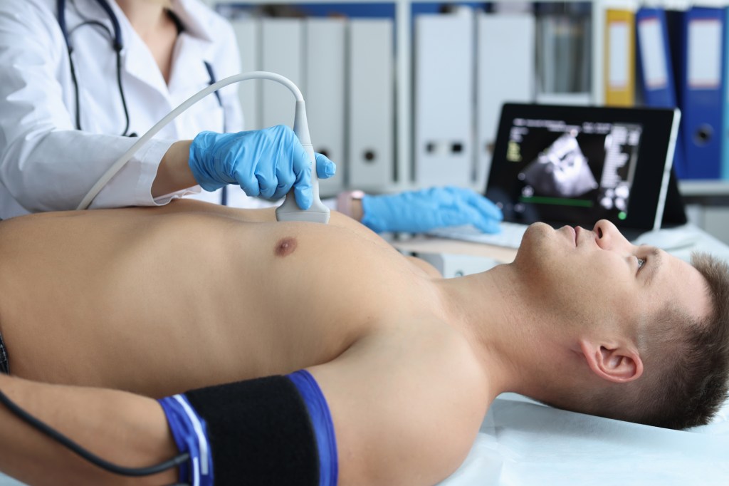
A sonographer places a hand-held wand (called a transducer) on the outside of your chest to send sound waves to your heart. These sound waves bounce off the different parts of your heart.

These “echoes” then appear as pictures on the sonographer’s computer screen. These pictures can also be saved for your cardiologist and physician to review later.

Preparing for a transthoracic echo

There’s not much you need to do to prepare for this type of echo. In general:

  • You don’t need to avoid eating or drinking before a transthoracic echo.
  • Take your medications as you usually do.
  • Wear anything you’d like.
  • Leave anything valuable at home. You’ll be given a storage locker to use during the test.

What to expect during a transthoracic echo

A transthoracic echo includes the following steps:

  1. You’ll be asked to remove your clothing from the waist up. You’ll put on a hospital gown.
  2. Your sonographer will place several electrodes on your chest. These are small, flat, sticky patches. The electrodes are attached to an electrocardiograph (EKG) monitor. The EKG records your heart’s electrical activity during the test.
  3. You’ll lie down on an exam table. Your sonographer will ask you to lie on your left side if possible.
  4. Your sonographer will place a sound-wave transducer (wand) on several areas of your chest. There’s a small amount of gel on the end of the wand, which won’t harm your skin. This gel helps produce clearer pictures.
  5. You may hear swishing sounds throughout the test. This is normal. It means you’re hearing blood flowing through your heart as the wand picks up the sound.
  6. Throughout the test, your sonographer may ask you to hold your breath for several seconds at a time. You may also need to move into a different position.

You should feel no major discomfort during the test. You may feel a coolness on your skin from the gel on the wand. You may also feel a slight pressure of the wand against your chest.

How is a transesophageal echocardiogram done?

A transesophageal echo takes pictures from inside your chest, rather than from the outside. It can show your heart and valves in greater detail than a transthoracic echo. That’s because your body’s bones and tissues aren’t in between the transducer and your heart.

For this test, the sonographer guides a small transducer down your throat and esophagus (food tube) using a long, flexible tube. This minimally invasive procedure may cause mild, temporary discomfort. But it has a low risk of serious problems.

This type of echo may be used:

  • When your provider needs a detailed look at your aorta or the back of your heart (especially your left atrium or left ventricle).
  • To check for blood clots.
  • To evaluate your mitral valve or aortic valve.
  • If you have obesity or lung disorders.
  • If a transthoracic echo isn’t possible for various other reasons.

Preparing for a transesophageal echo

As you prepare for your echo, tell your doctor if you have:

  • Problems with your esophagus, like a hiatal hernia.
  • Problems swallowing.
  • Sleep apnea.
  • IV drug use.

It’s also important to share if you take medication for:

  • Sleep issues.
  • Anxiety.
  • Pain.

Preparations for the day of your test include:

  • Find someone to drive you home from your test. This is because you’ll be sedated for the test, and you won’t be able to drive for 24 hours.
  • Don’t eat or drink anything for at least six hours before your test. Your provider may give you more specific instructions for eating and drinking. It’s essential you follow these closely.
  • Ask your provider about when to take your usual medications. You may be able to take them at your usual time with a small sip of water.
  • Ask your provider about when and how to take your diabetes medication, if this is relevant to you.
  • Plan to leave any valuable personal items at home. You’ll have access to a storage locker for your belongings during the test.
  • Plan to wear whatever’s most comfortable for you. You’ll change into a hospital gown before the echo begins.

What to expect during a transesophageal echo

A transesophageal echo includes the following steps:

  1. You’ll remove your clothing from the waist up and put on a hospital gown.
  2. Your provider will place electrodes (small stickers) on your chest. The electrodes allow your sonographer to monitor your heart’s electrical activity during the test.
  3. Your provider will place a blood pressure cuff on your arm and a pulse oximeter on your finger.
  4. You’ll gargle with a solution that numbs your throat. Your provider will also spray your throat with pain-relieving medication.
  5. To make you more comfortable, you’ll be hooked up to an IV and sedated. You’ll soon start to feel sleepy.
  6. You may have a tube in your nose that provides oxygen.
  7. You’ll lie on your left side on the exam table.
  8. The provider will insert an endoscope into your mouth. This is a long, thin, flexible tube that has a transducer on the tip. The tube travels into your throat and esophagus. It’s lubricated to help it slide down more easily. While this may feel uncomfortable, it won’t harm you. You may need to swallow to move the transducer into the right spot (just behind your heart).
  9. Your provider will take pictures. You won’t feel anything while this happens.
  10. After your provider has the pictures they need, they’ll remove the tube from your throat. You’ll soon be able to get dressed and get ready to leave.

How is an exercise stress echocardiogram done?

An exercise stress echo, sometimes simply called a stress echo, shows how your heart works when it’s taxed. The test resembles a traditional exercise stress test. A technician will monitor your heart rate and rhythm as well as your blood pressure (this is standard during a stress test). But they’ll also use echo imaging (which isn’t normally used during a stress test).

This test shows how well your heart can withstand activity. Your sonographer takes pictures before you start exercising and then right after you’re done.

In some cases, you won’t exercise. Instead, your provider will give you medication to make your heart work harder as if you were exercising. The goal is to force your heart to need more oxygen.

When your heart is under stress, your sonographer can see details they might not be able to see if you were lying on the exam table. These include problems with your coronary arteries or the lining of your heart.

Preparing for an exercise stress echo

Your provider will give you detailed instructions on how to prepare for your test. An exercise stress echo needs more preparation than other types of echo testing. These include:

  • Not eating or drinking anything except water for at least four hours before your test.
  • Not smoking on the day of your test.
  • Avoiding caffeine for 24 hours before your test. This includes caffeine in any form (coffee, tea, decaf drinks and some over-the-counter pain medicines).

Ask your provider when and how to take your usual medications. You may need to avoid taking certain heart medications on the day of your test. You may also need to change your dose of diabetes medication. Closely follow your provider’s guidance.

You won’t be sedated, but you still may want to ask someone to drive you to and from the appointment. You may feel tired after the test.

Plan to wear comfortable clothes and shoes. You’ll need to walk or ride a stationary bike during the test, so wear what feels good for you.

What to expect during an exercise stress echo

The exercise stress echo will include the following steps:

  1. Your sonographer will place electrodes (small stickers) on your chest. These stickers are hooked up to an EKG monitor to check your heart rate and rhythm during the test.
  2. Your provider will measure your heart rate, heart rhythm and blood pressure before you start moving.
  3. You’ll lie on an exam table so your sonographer can take pictures of your heart. They’ll place a hand-held wand (the kind usually used for echo tests) on the outside of your chest in various spots.
  4. Then, it’s time to start moving. You’ll walk on a treadmill or pedal on a stationary bike. The intensity will gradually increase. You’ll keep going until you’re exhausted. This usually takes seven to 12 minutes.
  5. As you’re exercising, a technician will ask how you’re feeling. Tell them any and all symptoms you notice. They’ll also watch your heart on the EKG monitor.
  6. You’ll stop moving, and you’ll have another echo test done.
  7. You’ll then do a short cool-down (slow walking or cycling), and your provider will monitor your vitals until they’re back to normal.

If you were given medication to stress your heart, the process will be a bit different. You won’t be on a treadmill or bike. Talk to your provider to learn what to expect and how you might feel during this type of test.

How do I get the results of my test?

After your cardiologist reviews your test, they’ll enter the results into your electronic medical record. Your primary care provider will have access to the results, too. You’ll discuss the results with one or both of these providers.

Ask any questions you’d like about the pictures and what they mean. Your provider will explain what the pictures show and whether you need follow-up tests or treatment.

What should you not do before an echocardiogram?

It depends on which type of echo you’re having done. Check with your provider to learn exactly what you should avoid. Things you may need to avoid before your echo include:

  • Eating or drinking.
  • Smoking or using any nicotine products.
  • Drinking coffee or anything with caffeine in it. This includes decaf drinks, which still contain a small amount of caffeine. It also includes over-the-counter medications that contain caffeine.

You may need to adjust your medication schedule before your echo. Don’t stop taking any medications or make any changes until you talk with your provider.

A note from QBan Health Care Services

An echocardiogram is an important test that can reveal a lot about your heart’s structure and function. If your provider recommends an echo for you, ask about what type you’ll be receiving and what you can expect. You may need more than one echo, or multiple tests with different techniques, so your provider can get enough details about your heart.

Ask your provider to explain the pictures to you and help you understand what they mean. Taking an active role in your diagnosis and care can help you feel comfortable with each step of the process.

SARCOPENIA

Sarcopenia is the age-related progressive loss of muscle mass and strength. The main symptom of the condition is muscle weakness. Sarcopenia is a type of muscle atrophy primarily caused by the natural aging process. Scientists believe being physically inactive and eating an unhealthy diet can contribute to the disease.

What is sarcopenia?

The medical definition of sarcopenia is the gradual loss of muscle mass, strength and function. The condition commonly affects the elderly population and is thought to occur due to aging. Sarcopenia can greatly impact your quality of life by reducing your ability to perform daily tasks. It can lead to the loss of your independence and the need for long-term care.

Sarcopenia affects your musculoskeletal system and is a major factor in increased frailty, falls and fractures. These conditions can lead to hospitalizations and surgeries, which increase the risk of complications including death.

Sarcopenia can also affect people with a high body mass index (BMI), in a condition called sarcopenic obesity. People with obesity and sarcopenia have a greater risk for complications than with obesity or sarcopenia alone.

Who does sarcopenia affect?

Sarcopenia most commonly affects people ages 60 and older. The rates increase with age. The disease affects both sexes equally. Studies on affected ethnicities are inconsistent. The rates of the condition increase in people with chronic disease.

How common is sarcopenia?

Studies are inconsistent, and many people don’t receive a diagnosis or treatment for sarcopenia. But rates of the condition range from 5% to 13% in people ages 60 and older. The estimates increase to 11% to 50% in people ages 80 and older.

How does sarcopenia affect my body?

A decrease in both the number and size of your muscle fibers causes your muscles to thin (muscle atrophy).

As you age, your body goes through certain changes that play a major factor in developing sarcopenia. For instance, your body doesn’t produce the same amount of proteins your muscles need to grow. When this happens, your muscle cells get smaller.

In addition, as you grow older, changes in certain hormones — like testosterone and insulin-like growth factor (IGF-1) — affect your muscle fibers. This can lead to sarcopenia.

What are the symptoms of sarcopenia?

The most common symptom of sarcopenia is muscle weakness. Other symptoms may include:

  • Loss of stamina.
  • Difficulty performing daily activities.
  • Walking slowly.
  • Trouble climbing stairs.
  • Poor balance and falls.
  • Decrease in muscle size.

What causes sarcopenia?

The most common cause of sarcopenia is the natural aging process. You gradually begin losing muscle mass and strength sometime in your 30s or 40s. This process picks up between the ages of 65 and 80. Rates vary, but you may lose as much as 8% of your muscle mass each decade. Everyone loses muscle mass over time, but people with sarcopenia lose it more quickly.

Although aging tends to be the dominant factor, researchers have discovered other possible risk factors for sarcopenia. These may include:

  • Physical inactivity.
  • Obesity.
  • Chronic diseases such as chronic obstructive pulmonary disease (COPD), kidney disease, diabetes, cancer and HIV.
  • Rheumatoid arthritis.
  • Insulin resistance.
  • Reduction in hormone levels.
  • Malnutrition or inadequate protein intake.
  • Decrease in your ability to convert protein to energy.
  • Decline in the number of nerve cells that send messages from your brain to your muscles telling them to move.

How is sarcopenia diagnosed?

Your healthcare provider may diagnose sarcopenia after performing a physical exam and asking you about your symptoms. You may complete a questionnaire based on your self-reported symptoms called the SARC-F. SARC-F stands for:

  • S — Strength.
  • A — Assistance with walking.
  • R — Rising from a chair.
  • C — Climbing stairs.
  • F — Falls.

You score each factor with a number between 0 and 2. The highest maximum SARC-F score is 10. A SARC-F score of 4 or more warrants more testing.

What tests will be done to diagnose sarcopenia?

There is no single test that can diagnose sarcopenia. Your healthcare provider may recommend several tests to diagnose and then determine the severity of sarcopenia.

Muscle strength tests

  • Handgrip test: Handgrip strength draws a parallel to the strength in your other muscles. Providers use it to identify shortages in overall muscle strength.
  • Chair stand test: Providers use the chair stand test to measure your leg muscle strength, especially your quadriceps. The chair stand test measures the number of times you can stand and sit from a chair without the use of your arms in 30 seconds.
  • Walking speed test: The walking (gait) speed test measures the time it takes for you to travel 4 meters (about 13 feet) at your usual walking pace.
  • Short physical performance battery (SPPB): With the SPPB test, you take three timed tasks: chair stand test, standing balance test and walking speed test.
  • Timed-up and go test (TUG): The TUG test measures the time it takes for you to rise from a chair, walk 3 meters (about 10 feet) away from the chair, walk 3 meters back to the chair, and sit back down in the chair.

Imaging tests for measurement of muscle mass

  • Dual-energy X-ray absorptiometry (DEXA or DXA): This type of imaging test uses low-energy X-rays to measure your muscle mass, fat mass and bone density.
  • Bioelectrical impedance analysis (BIA): The BIA test is less expensive and more widely available than DEXA. It measures your body fat in relation to your lean body mass.

How is sarcopenia treated?

Treatment for sarcopenia typically includes lifestyle changes. These modifications to your lifestyle behaviors can treat and help reverse sarcopenia.

  • Physical activity: Your healthcare provider may recommend progressive resistance-based strength training. This type of exercise can help improve your strength and reverse your muscle loss.
  • Healthy diet: When paired with regular exercise, eating a healthy diet can also help reverse the effects of sarcopenia. It’s especially important to increase your protein intake through food or supplements.

What medications are used to treat sarcopenia?

Researchers are studying the possibility of using hormone supplements to increase muscle mass. But there aren’t currently any FDA-approved medications to treat sarcopenia.

How can I prevent sarcopenia?

You may not be able to completely prevent sarcopenia since the condition happens as part of the natural aging process. But you can take steps to slow the progression of the disease. These include:

  • Make healthy food choices: Maintain a healthy diet that includes high-quality proteins. Aim for 20 to 35 grams of protein in each meal.
  • Exercise: Maintain a physically active lifestyle that includes exercises such as resistance training.
  • Routine physicals: See your healthcare provider regularly, and let them know about any changes in your health.

What can I expect if I have sarcopenia?

The outlook for sarcopenia primarily depends on your age. Rates of the condition increase as you grow older. In addition, the outlook for the condition varies based on your health and lifestyle.

The disease can greatly affect your quality of life. You may be able to reverse the effects of the condition with lifestyle changes. If you don’t make recommended changes, the disease will continue to weaken your muscles. Over time, you may need full-time care to live your life.

Is sarcopenia a disease?

In 2016, the Centers for Disease Control and Prevention (CDC) declared sarcopenia a specific disease by creating an International Classification of Disease (ICD) 10 code for the condition. This designation made sarcopenia a reportable disease by healthcare providers. Therefore, it increased the diagnosis and treatment of the disease. This label helps distinguish sarcopenia from similar diseases and allows researchers to begin collecting valuable data about the condition.

What’s the difference between sarcopenia and muscle atrophy?

Sarcopenia is a type of muscle atrophy that specifically affects people as they grow older. Muscle atrophy is the loss of muscle tissue. The two conditions share common features of muscle loss, but the processes behind them are different. A decrease in the size and number of your muscle fibers causes sarcopenia. With muscle atrophy, there’s a reduction in the size of the fibers, but the amount of fibers stays the same.

A note from QBan Health Care Services

Everyone experiences some amount of muscle loss as they age. But with sarcopenia, this muscle loss happens faster. The good news is, there are ways to treat and even reverse the effects of the condition. If you’ve experienced muscle weakness, loss of endurance or any other symptoms of sarcopenia, call your healthcare provider. They can diagnose the condition and develop a treatment plan for you to revert the muscle loss and improve your condition.

OSTEOPOROSIS

Osteoporosis silently weakens your bones, which can make you more likely to experience a bone fracture (broken bone). You can prevent bone density loss with treatments and exercise. Ask your provider about a bone density test if you’re over 65 or have a family history of osteoporosis.

What is osteoporosis?

Osteoporosis is a disease that weakens your bones. It makes your bones thinner and less dense than they should be. People with osteoporosis are much more likely to experience broken bones (bone fractures).

Your bones are usually dense and strong enough to support your weight and absorb most kinds of impacts. As you age, your bones naturally lose some of their density and their ability to regrow (remodel) themselves. If you have osteoporosis your bones are much more fragile than they should be, and are much weaker.

Most people don’t know they have osteoporosis until it causes them to break a bone. Osteoporosis can make any of your bones more likely to break, but the most commonly affected bones include your:

The sooner a healthcare provider diagnoses osteoporosis, the less likely you are to experience bone fractures. Ask a healthcare provider about checking your bone density, especially if you’re over 65, have had a bone fracture after age 50, or someone in your biological family has osteoporosis.

How common is osteoporosis?

More than 50 million people in the U.S. live with osteoporosis.

Osteoporosis is common in people over 50. Experts estimate that half of all people assigned female at birth and 1 in 4 people assigned male at birth over 50 have osteoporosis.

Studies have found that 1 in 3 adults over 50 who don’t have osteoporosis yet have some degree of reduced bone density (osteopenia). People with osteopenia have early signs of osteoporosis. If it’s not treated, osteopenia can become osteoporosis.

What are osteoporosis symptoms?

Osteoporosis doesn’t have symptoms the way lots of other health conditions do. That’s why healthcare providers sometimes call it a silent disease.

You won’t feel or notice anything that signals you might have osteoporosis. You won’t have a headache, fever or stomachache that lets you know something in your body is wrong.

The most common “symptom” is suddenly breaking a bone, especially after a small fall or minor accident that usually wouldn’t hurt you.

Even though osteoporosis doesn’t directly cause symptoms, you might notice a few changes in your body that can mean your bones are losing strength or density. These warning signs of osteoporosis can include:

  • Losing an inch or more of your height.
  • Changes in your natural posture (stooping or bending forward more).
  • Shortness of breath (if disks in your spine are compressed enough to reduce your lung capacity).
  • Lower back pain (pain in your lumbar spine).

It might be hard to notice changes in your own physical appearance. A loved one may be more likely to see changes in your body (especially your height or posture). People sometimes joke about older adults “shrinking” as they age, but this can be a sign that you should visit a healthcare provider for a bone density test.

What causes osteoporosis?

Osteoporosis happens as you get older and your bones lose their ability to regrow and reform themselves.

Your bones are living tissue like any other part of your body. It might not seem like it, but they’re constantly replacing their own cells and tissue throughout your life. Up until about age 30, your body naturally builds more bone than you lose. After age 35, bone breakdown happens faster than your body can replace it, which causes a gradual loss of bone mass.

If you have osteoporosis, you lose bone mass at a greater rate. People in postmenopause lose bone mass even faster.

Osteoporosis risk factors

Anyone can develop osteoporosis. Some groups of people are more likely to experience it, including:

  • Anyone over 50.
  • People assigned female at birth (AFAB), especially people AFAB in postmenopause.
  • People with a family history (if someone in your biological family has osteoporosis).
  • People who are naturally thin or who have “smaller frames.” People with thinner statures often have less natural bone mass, so any losses can affect them more.
  • People who smoke or use tobacco products.

Some health conditions can make you more likely to develop osteoporosis, including:

Some medications or surgical procedures can increase your risk of osteoporosis:

Certain aspects of your diet and exercise routine can make you more likely to develop osteoporosis, including:

  • Not getting enough calcium or vitamin D in your diet.
  • Not getting enough physical exercise.
  • Regularly drinking alcohol (more than two drinks per day).

How is osteoporosis diagnosed?

A healthcare provider will diagnose osteoporosis with a bone density test. A bone density test is an imaging test that measures the strength of your bones. It uses X-rays to measure how much calcium and other minerals are in your bones.

Healthcare providers sometimes refer to bone density tests as DEXA scans, DXA scans or bone density scans. All of these are different names that refer to the same test.

A bone density test uses low levels of X-rays to measure the density and mineral content of your bones. It’s similar to a typical X-ray. It’s an outpatient procedure, which means you won’t have to stay in the hospital. You can go home as soon as you finish your test. There are no needles or injections in this test.

Checking for changes in your bone density is the best way to catch osteoporosis before it causes a bone fracture. Your provider might suggest you get regular bone density tests if you have a family history of osteoporosis, if you’re over 50, or you have osteopenia.

How is osteoporosis treated?

Your healthcare provider will suggest a combination of treatments that slow down your bone loss and strengthen your existing bone tissue. The most important part of treating osteoporosis is preventing bone fractures.

The most common osteoporosis treatments include:

  • Exercise: Regular exercise can strengthen your bones (and all the tissue connected to them, like your muscles, tendons and ligaments). Your provider might suggest weight-bearing exercise to strengthen your muscles and train your balance. Exercises that make your body work against gravity like walking, yoga, Pilates and tai chi can improve your strength and balance without putting too much stress on your bones. You might need to work with a physical therapist to find exercises and movements that are right for you.
  • Vitamin and mineral supplements: You might need over-the-counter or prescription calcium or vitamin D supplements. Your provider will tell you which type you need, how often you should take them and which dosage you’ll need.
  • Medications for osteoporosis: Your provider will tell you which prescriptions will work best for you and your body. Some of the most common medications providers use to treat osteoporosis include hormone therapies like replacement estrogen or testosterone and bisphosphonates. People with severe osteoporosis or a high risk of fractures might need medications, including parathyroid hormone (PTH) analogs, denosumab and romosozumab. These medications are usually given as injections.

How can I lower my risk of osteoporosis?

Exercise and making sure you get enough calcium and vitamin D in your diet are usually all you’ll need to prevent osteoporosis. Your provider will help you find a combination of treatments that’s best for you and your bone health.

Follow these general safety tips to reduce your risk of an injury:

  • Always wear your seatbelt.
  • Wear the right protective equipment for all activities and sports.
  • Make sure your home and workspace are free from clutter that could trip you or others.
  • Always use the proper tools or equipment at home to reach things. Never stand on chairs, tables or countertops.
  • Follow a diet and exercise plan that’s healthy for you.
  • Use a cane or walker if you have difficulty walking or have an increased risk for falls.

What can I expect if I have osteoporosis?

You should expect to manage osteoporosis for a long time, usually the rest of your life. You’ll need regular appointments with a healthcare provider and bone density tests. Your provider will monitor any changes in your bone density and will adjust your treatments as needed.

How do I take care of myself?

Following a diet and exercise plan that’s healthy for you will help you maintain your bone (and overall) health. See a healthcare provider for regular checkups. They’ll also help catch any issues or symptoms that affect your bones as soon as possible.

Talk to your provider about a bone density test if you’re over 65 or have a family history of osteoporosis.

When should I see my healthcare provider?

Visit a healthcare provider if you notice any changes in your body that might be osteoporosis warning signs. Tell your provider about any other symptoms you’re experiencing, especially if you have bone pain or trouble moving.

When should I go to the ER?

Go to the emergency room if you think you have a broken bone or if you experience any of the following symptoms:

  • Intense pain.
  • You can’t move a part of your body.
  • A part of your body is noticeably different looking or out of its usual place.
  • You can see your bone through your skin.
  • Swelling.
  • New bruising that appears at the same time as any of these other symptoms.

What questions should I ask my healthcare provider?

  • What is my risk for developing osteoporosis?
  • How often will I need bone density tests?
  • Which treatments will I need?
  • What are some good exercises I can do to help strengthen my bones?
  • Will I need physical therapy?

What is the life expectancy of someone with osteoporosis?

Osteoporosis itself isn’t fatal and won’t change your life expectancy (how long you’ll live). But it can make you more likely to experience a bone fracture (and can increase your risk of more severe breaks or complications from a fracture). Some studies have found that hip fractures in adults older than 65 lead to reduced mobility and an earlier death.

Talk to your healthcare provider if you’re worried about your risk of falls or bone fractures. They’ll help you stay safe and healthy.

A note from QBan Health Care Services

Osteoporosis makes your bones thinner and weaker than they should be. It can be dangerous because it makes you more likely to experience a bone fracture. Lots of people don’t even know they have osteoporosis until it causes a broken bone.

The best way to prevent bone fractures is catching osteoporosis before it can hurt you. Visit a healthcare provider for regular checkups. Ask them when you’ll need bone density tests and how often you should have follow-up tests to monitor your bone health.

TRANSVAGINAL ULTRASOUND

A transvaginal ultrasound is a relatively quick, painless imaging procedure that provides a more detailed view of your pelvic organs than a traditional abdominal ultrasound provides. Your provider may order this imaging procedure to explore what’s causing your symptoms, diagnose a condition, monitor your pregnancy or plan for surgery. It’s one of the most common, most useful diagnostic tools your provider can use to provide you care.

What is a transvaginal ultrasound?

A transvaginal ultrasound is an imaging procedure that allows your provider to see your pelvic cavity and the organs inside your pelvis. These organs include your cervix, uterus, fallopian tubes and ovaries. A transvaginal ultrasound can show abnormal structures or growths in your pelvic area that may indicate a condition or disease. Your provider may also order a transvaginal ultrasound to confirm or monitor your pregnancy.

What is the difference between an ultrasound and a transvaginal ultrasound?

Transvaginal ultrasounds are sometimes called “endovaginal ultrasounds” because the device that records images of your pelvic cavity (transducer) is inserted inside your vagina. The process is different from a traditional abdominal ultrasound, where your provider moves the transducer across your belly to record images.

A transvaginal ultrasound provides a more detailed visual of your organs and the soft tissue inside your pelvic cavity than an abdominal ultrasound. But, an abdominal ultrasound is an option if a transvaginal ultrasound cannot be done. For instance, if you’re pregnant and the placenta (the organ that provides nutrients to your baby) is near your cervix, a transvaginal ultrasound may cause unnecessary bleeding.

When is a transvaginal ultrasound performed?

There aren’t many restrictions or limiting factors when your provider can perform a transvaginal ultrasound. For instance, your provider may order this procedure regardless of whether or not you’re menstruating or pregnant.

There are limitations If your provider recommends a special kind of transvaginal ultrasound called saline infusion sonohysterography, or a sonohysterogram. A sonohysterogram is when a small amount of fluid is used to distend the cavity of the uterus to allow a good imaging view of the lining. You shouldn’t have a sonohysterogram if you’re pregnant  or have pelvic inflammatory disease (PID).

When would a transvaginal ultrasound be needed?

Your provider may order a transvaginal ultrasound to diagnose conditions causing unpleasant symptoms, like pelvic pain or abnormal bleeding. Transvaginal ultrasounds can provide further information about abnormalities discovered during a pelvic exam that can lead to a diagnosis. Transvaginal ultrasounds may also be used to monitor your pregnancy.

A transvaginal ultrasound allows your provider to identify:

Your provider may order a transvaginal during pregnancy, especially during the first trimester (weeks one to 12). Your provider may order a transvaginal ultrasound to:

  • Confirm your pregnancy.
  • Determine how far along you are in your pregnancy.
  • Monitor your baby’s heartbeat.
  • Identify warning signs of a miscarriage or early delivery.
  • Identify conditions that will affect your pregnancy plan, like having a low-lying placenta (placenta previa).

A transvaginal ultrasound can also help your provider plan for surgery.

Who performs a transvaginal ultrasound?

Your provider or a trained specialist called a sonographer, or ultrasound technician may perform the procedure. In some instances, a trained radiologist may perform the procedure and share the results with your provider.

How does a transvaginal ultrasound work?

A transvaginal ultrasound uses sound waves to record your pelvic cavity and organs and project these images onto a screen. A wand-like instrument called a transducer is inserted into your vagina, where it releases sound waves that bounce off the various structures inside your pelvis. The sound waves travel back to the transducer, where they’re converted into electrical signals. These signals project a real-time visual image of your pelvic organs onto a screen that the technician performing the procedure can view.

The ultrasound captures still images of the visuals on screen, too, so that your provider can examine them later. The image produced during an ultrasound is called a “sonogram.”

How long does a transvaginal ultrasound take?

A transvaginal ultrasound can take anywhere from 15 minutes to an hour.

How do I prepare for a transvaginal ultrasound?

Transvaginal ultrasounds don’t require much preparation. The procedure is quick and relatively painless, with little risk of side effects. You shouldn’t have to plan for someone to drive you home or to work afterward.

To prepare:

  • Wear clothes that you can slip out of easily. You will have to remove your pants and underwear, and you may have to wear a gown.
  • Remove your tampon before the procedure if you’re on your period.
  • Follow your provider’s instructions about when to drink fluids and go to the bathroom. Having a full bladder affects the way your organs appear on an ultrasound. You may need to arrive at your appointment with a bladder that’s empty, full or partially full.

Do I need to shave for a transvaginal ultrasound?

No. Having pubic hair won’t prevent you from having an ultrasound. Groom to your comfort before the procedure.

What should I expect during a transvaginal ultrasound?

Your ultrasound will occur in an ultrasound room in a hospital, private radiology practice or clinic for obstetric and gynecological imaging. In some instances, your provider may order an abdominal ultrasound before your transvaginal ultrasound. Both imaging procedures together provide a more comprehensive view of your pelvic organs that may be needed, depending on your symptoms.

  1. You’ll lie on an examination table as if you were having a pelvic exam — with your knees bent, and your feet possibly in stirrups.
  2. Your provider will place a condom and a warm lubricating gel on the transducer and gently insert it inside your vagina.
  3. Once it’s inside your body, the transducer releases sound waves that record pictures of your pelvic organs. These images get projected onto a screen. The technician performing the ultrasound may ask that you lie still or shift your body so that the transducer can record your pelvic cavity from different angles.

The technician may perform additional steps if you’re having saline-infusion sonography, or a sonohysterogram.

Once enough images are taken for a thorough analysis, the technician will remove the transducer.

Is a transvaginal ultrasound painful?

No. The transducer is designed to curve to your vagina’s shape so that the procedure is as painless as possible. Also, the lubricating gel placed on the transducer allows for gentle insertion. Still, you may feel some discomfort or pressure when the technician inserts the transducer into your vagina. The process may feel similar to a Pap smear, when your provider uses a speculum to widen the opening in order to access your cervix.

What are the risks of a transvaginal ultrasound?

Transvaginal ultrasounds are safe. The sound technology used to record images of your pelvis doesn’t pose risks to you or your fetus if you’re pregnant. You may feel slight discomfort or pressure from the transducer. And you may have a slight vaginal discharge from the lubricating gel used during the procedure that should disappear after 24 hours.

What type of results do you get after a transvaginal ultrasound?

If your provider performs the procedure, they may discuss your results with you that same day. Or, they may send the images to a certified physician sonologist for analysis. The sonologist will examine your ultrasound images and type up a report communicating their findings to your provider.

Your test results may provide enough information for a definitive diagnosis. Alternatively, your provider may request additional testing if there’s uncertainty. Ask your provider about how your results will affect your care plan.

A note from QBan Health Care Services

A transvaginal ultrasound is a common procedure that can help your provider identify what’s causing unpleasant symptoms you may be experiencing. It requires little effort on your part while providing helpful information to your provider about conditions that require a range of treatment options. Speak to your provider about how having a transvaginal ultrasound will inform the next steps of your care.