Whooping cough (pertussis) is a very contagious respiratory infection that causes a distinctive “whooping” cough. The condition can cause bouts of repeated, violent coughing that may come and go and last for weeks or months. Pertussis can cause severe, life-threatening illness, especially in babies. The pertussis vaccine can help prevent it.
What is whooping cough?
Whooping cough, also called pertussis, is a very contagious upper respiratory infection. It usually gives you lengthy and repeated bouts of coughing. These coughing episodes can continue for weeks or even months after you first develop symptoms of the illness.
Instead of coughing spells, babies with pertussis may have breathing difficulties. This includes conditions such as apnea, when there are pauses in their breathing. Whooping cough can cause serious, life-threatening complications in babies. About one-third of all infants (babies younger than 1 year old) who get whooping cough need treatment at a hospital.
The whooping cough vaccine can help prevent the infection.
What does whooping cough sound like?
Prolonged coughing causes air to be expelled from your lungs. When you gasp for air quickly and deeply after a coughing fit, a whooping cough sound might accompany the inhalation of air. This sound is a loud, high-pitched “whooping” noise. That’s where pertussis gets its name. However, someone may still have the infection without making the noise.
Who does whooping cough affect?
Pertussis can affect anyone, but it most often occurs in babies, children and adolescents. Babies are especially vulnerable to infection because they can’t receive the pertussis vaccine until they’re at least 2 months old. They can catch whooping cough from their parents, adult caregivers or other children.
Can adults get whooping cough?
Yes. But whooping cough in adults is generally milder than in babies and children. This is especially true for adults who’ve received the whooping cough vaccine. The infection may seem more like the common cold. The “whoop” may not be there in people with milder illnesses.
However, adults can develop serious cases of whooping cough, especially if they haven’t received the pertussis vaccine. They may have long-lasting coughing fits that keep them awake all night. People who’ve experienced these coughing bouts say it’s the worst cough of their lives. It can also cause major interruptions in your daily life and serious complications.
How common is pertussis?
Before the development of the pertussis vaccine, there were hundreds of thousands of cases of whooping cough each year in the U.S.
Today, case numbers have dropped significantly. According to the Centers for Disease Control and Prevention (CDC), there were about 1,600 reported cases of pertussis in the U.S. in 2021.
Every few years, outbreaks occur, and there are peaks in reported cases. In addition, whooping cough continues to be a global endemic. According to the World Health Organization (WHO), there were more than 151,000 cases of pertussis worldwide in 2018.
What are the symptoms of whooping cough?
Early pertussis symptoms may resemble those of the common cold. These symptoms may persist for one to two weeks and may include:
- Slight fever.
- Mild or occasional coughing.
- Runny nose.
- A pause in breathing in babies (apnea).
Whooping cough symptoms after the first or second week have passed usually include:
- Prolonged, repeated or violent coughing episodes (paroxysms) that recur intermittently for up to 10 weeks or more.
- Whooping sound when inhaling after the coughing stops.
- Vomiting.
- Exhaustion due to prolonged coughing.
Symptoms of pertussis begin to lessen after four weeks, although bouts of coughing can recur for months after symptoms start.
What causes whooping cough?
A type of bacteria called Bordetella pertussis causes whooping cough. The condition starts when the bacteria enter your respiratory system. They attach to the tiny, hair-like extensions (cilia) on the lining of your respiratory tract. Then, they release poisons (toxins), which damage your cilia and cause your airways to swell. This swelling causes the secretions of your mucus to increase, which causes severe coughing.
Is whooping cough contagious?
Yes. When you cough or sneeze, you can spread tiny respiratory droplets containing the bacteria into the air. People around you may inhale these bacteria-containing droplets and get infected. You may be contagious even before symptoms appear and remain contagious for up to two weeks after coughing starts.
How is whooping cough diagnosed?
A healthcare provider will perform a physical examination and ask questions about your symptoms. They may use a cotton swab to take a sample of the mucus inside your nose. They can also collect a sample by filling a syringe with saline fluid and flushing it through your nose and the back of your throat.
The provider will send the samples to a lab where a technician will analyze them for the presence of Bordetella bacteria. The provider may also request blood tests to confirm the presence of the bacteria.
What is the treatment for whooping cough?
Whooping cough treatment should begin as soon as possible after diagnosis. A provider will prescribe antibiotics to help prevent the spread of the disease. But antibiotics can’t prevent or treat your cough. Cough syrups and other medicines can’t relieve your coughing spells, so you’ll need to use other forms of home treatment to manage your symptoms.
If you’ve been in close contact with someone infected with pertussis, you should start treating whooping cough within three weeks of exposure. A provider will likely recommend antibiotics as well.
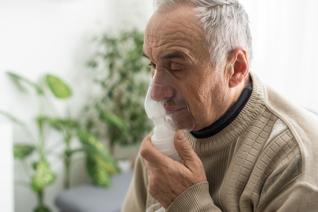
If your baby develops the condition, they may need pertussis treatment in the hospital. Whooping cough can cause life-threatening complications, such as pneumonia or breathing problems. To treat the infection and prevent complications, a healthcare provider will:
- Keep your baby’s breathing passages clear. They may have to suction out mucus.
- Monitor your baby’s breathing. They’ll give them oxygen, if necessary.
- Prevent or treat dehydration. Your baby may need an IV to receive fluids.
How can I prevent whooping cough?
Getting the whooping cough vaccine is the best way to prevent it getting whooping cough. Healthcare providers recommend that all children receive the diphtheria-tetanus-pertussis (DTaP) vaccine. This is a combination vaccine that also protects them against diphtheria and tetanus. It’s safe and effective.
Children should receive five injections of the DTaP vaccine, according to the following recommended vaccination schedule:
- First dose: 2 months old.
- Second dose: 4 months old.
- Third dose: 6 months old.
- Fourth dose: Between 15 and 18 months old.
- Fifth dose: Between 4 and 6 years old.
Healthcare providers recommend a booster vaccine for adults because they’re the most likely source of pertussis infection in infants. It’s important that all adults caring for infants receive a booster, as adults’ immunity to pertussis wanes as they get older. Adults who have a whooping cough infection may be told they have bronchitis or a sinus infection, and may unknowingly infect susceptible infants who are at risk for more serious complications.
Adults between the ages of 19 and 64 should receive a one-time booster pertussis vaccine called the Tdap vaccine.
Adults older than 64 years old should also receive a booster vaccine if they’ll come into close contact with babies younger than 12 months old. Pregnant people should receive a Tdap injection during the third trimester (between the 27th and 36th week) of their pregnancies. They must have a Tdap injection during each pregnancy.
The vaccine helps provide babies with short-term protection against whooping cough. It may also protect them against serious complications associated with the infection. If you’re pregnant, you should also make sure family members and caregivers take pertussis precautions by getting vaccinated.
What can I expect if I have whooping cough?
Pertussis may initially look like the common cold. But eventually, the telltale sign of the condition — the whooping cough sound — may develop. With prompt diagnosis, you can start on antibiotics that can help prevent the spread of the infection. But antibiotics won’t treat the cough. Your coughing episodes may last for weeks or even months. Whooping cough is more serious in babies and children, and serious complications can develop.
What are the possible complications of whooping cough?
Whooping cough can lead to serious, life-threatening complications, especially in babies younger than 1 year old. Approximately one-third of infants will need care in the hospital. Of those babies, about:
- 2 in 3 (68%) will develop pauses in their breathing called apnea.
- 1 in 5 (22%) will develop pneumonia.
- 1 in 50 (2%) will have violent, uncontrollable episodes of shaking (convulsions).
- 1 in 150 (0.6%) will develop a brain disease called encephalopathy.
- 1 in 100 (1%) will die.
Adults can also develop complications, but they’re usually less severe. If you have a persistent, violent cough, you may:
- Pass out.
- Break (fracture) a rib.
- Lose your bladder control.
- Lose weight unintentionally.
How do I take care of myself?
If you have whooping cough, there are several steps you can take to help manage your symptoms at home:
- Get plenty of rest.
- Drink lots of fluids to prevent dehydration.
- Use a cool-mist humidifier to soothe your lungs and loosen mucus in your respiratory tract.
- Take the antibiotics your provider prescribed as directed.
- Don’t take cough medicine unless recommended by your provider.
- Keep your home free from irritants such as smoke and dust that may trigger coughing fits.
When should I see my healthcare provider?
If you develop any signs of dehydration, you should seek advice from your healthcare provider immediately. Symptoms of dehydration may include:
- Dry mouth.
- Excessive thirst.
- Fatigue.
- Peeing less (decreased urination).
- Muscle weakness.
- Headache.
- Dizziness or lightheadedness.
A note from QBan Health Care Services
Whooping cough (pertussis) is a respiratory infection that can cause severe, prolonged coughing episodes or breathing difficulties. It can cause life-threatening illnesses, especially in babies. If you or your child develop a loud, “whooping” cough or you notice your baby is having trouble breathing, seek treatment right away. And if you haven’t already, get the pertussis vaccine to prevent infection.