SCLERODERMA

Scleroderma makes your body produce too much collagen, a protein that you need for healthy skin and tissue. It’s an autoimmune condition, which means your immune system attacks your body instead of protecting it. Scleroderma can cause lots of symptoms and affect tissue throughout your body. It can also lead to life-threatening complications.

What is scleroderma?

Scleroderma is a rare condition that makes your body produce tissue that’s thicker than it should be. Scleroderma usually affects your skin, but can cause symptoms in any tissue throughout your body.

Scleroderma is an autoimmune disorder. Autoimmune disorders happen when your immune system accidentally attacks your body instead of protecting it. Experts don’t know why your immune system turns on you. It’s like it can no longer tell the difference between what’s healthy and what’s not — between what’s you and what’s an invader like bacteria or a virus.

If you have scleroderma, your immune system triggers your body’s cells to produce too much collagen (a protein). Your body needs collagen to have strong, healthy connective tissue to support your organs and hold parts of your body in place. But when you produce too much of it, your skin and other tissue can be thicker and more fibrous than they should be.

Scleroderma is a chronic condition, which means you’ll need to manage your symptoms for a long time (maybe the rest of your life). It can also cause life-threatening complications if it affects tissue in your organs. Call 911 (or your local emergency services number) or go to the emergency room if you feel like you’re having a heart attack, can’t breathe or can’t swallow.

Visit a healthcare provider if you’re experiencing symptoms like pain and stiffness in your joints, especially if you notice thickened skin around your fingers and toes.

Types of scleroderma

Healthcare providers classify scleroderma into two main types:

Localized scleroderma: Localized means concentrated in one area. Localized scleroderma only affects one part of your body (usually your skin). It causes thick patches or streaks on your skin that feel waxy. Localized scleroderma can get better (resolve) on its own. It usually doesn’t spread to other parts of your body.

Systemic sclerosis: Systemic sclerosis can affect other organs, in addition to your skin. It can affect parts of your respiratory system (the organs that help you breathe and smell) and your digestive system (the organs that help you turn foods and drinks into energy). Scleroderma is more likely to cause serious complications if it affects your ability to breathe or process nutrition. It can be fatal. Systemic sclerosis has three subtypes — diffuse, limited and sine sclerosis.

Diffuse sclerosis

Diffuse means spread out widely. Diffuse sclerosis causes thickened skin over larger areas at once, including your:

  • Chest.
  • Abdomen.
  • Thighs.
  • Arms.
  • Legs.
  • Face.

It can also affect multiple organs at once, including your:

Limited sclerosis (CREST syndrome)

Healthcare providers usually refer to limited scleroderma with the acronym CREST syndrome. Each letter in CREST stands for a symptom it causes:

  • Calcinosis (extra calcium deposits in your skin).
  • Raynaud’s syndrome (color changes and numbness in your fingertips and toes).
  • Esophageal dysfunction (difficulty swallowing and acid reflux).
  • Sclerodactyly (tight skin on your fingers).
  • Telangiectasias (red or discolored spots on your skin).

Sine sclerosis

Sine sclerosis causes limited sclerosis symptoms, but doesn’t affect your skin. You may have any CREST syndrome symptoms but not experience any thickened skin.

How common is scleroderma?

Scleroderma is rare. Experts estimate that all types of scleroderma affect around 250 out of every 1 million people in the U.S. Around 100,000 people in the U.S. have systemic scleroderma.

What are scleroderma symptoms?

Some people with early scleroderma don’t have any symptoms. The most common scleroderma symptom is having patches or streaks of thickened, waxy skin. Other common symptoms include:
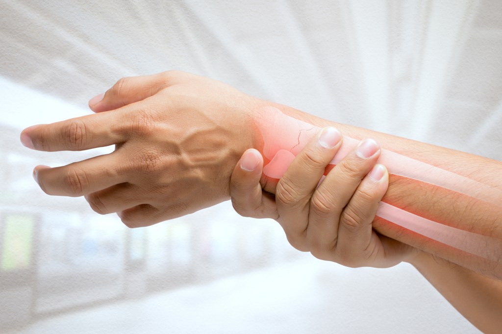
Which other symptoms you experience (and where they affect you) depends on which type of scleroderma you have.

Localized scleroderma symptoms

People with localized scleroderma usually only experience skin thickening. The thickened skin can be isolated to one specific area or appear in patches. It can affect skin on your:

  • Chest.
  • Abdomen (the area around your stomach).
  • Arms and legs (your limbs).
  • Hands and fingers.
  • Feet and toes.

It’s rare for localized scleroderma to affect your internal organs.

Systemic sclerosis symptoms

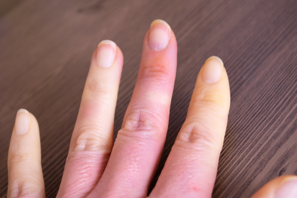
Systemic scleroderma can cause lots of symptoms. It causes thickened skin, usually in larger areas and patches, including on your face and hands. Thick skin usually appears on your fingers or toes and then spreads toward the center of your body. If you have Raynaud’s syndrome, the skin on your affected fingers and toes may change color when they’re exposed to cold (usually white, reddish or purple).

Systemic sclerosis can also cause symptoms in other organs and tissue, like your:

What causes scleroderma?

Experts don’t know for sure what causes scleroderma.

Some studies have found that it can run in families (meaning biological parents can pass it on to their children), but this is rare enough that there’s no definite proof it’s a genetic disorder.

Scleroderma risk factors

Anyone can develop scleroderma, but some groups of people have a higher risk:

  • Females are four times more likely than males to develop scleroderma.
  • People 30 – 50 years old — it’s rare for people younger than 30 to have scleroderma.
  • Black people are more likely to have scleroderma, usually develop it earlier and are more likely to experience symptoms that affect their lungs. They also usually have more severe skin symptoms.

What complications can scleroderma cause?

People with scleroderma are much more likely to have two other conditions: Raynaud’s syndrome and Sjögren’s syndrome.

Reynaud’s syndrome affects small blood vessels in your fingers and toes (your digits). People with it have episodes of symptoms (sometimes called attacks). It makes blood vessels in your digits suddenly tighten (contract) more than they should. This can make the skin in your affected digits turn pale or lighter than your natural skin tone. They might also look bluish.

Sjögren’s syndrome makes your body produce less moisture in certain glands — usually the salivary glands in your mouth and the glands in your eyes that produce tears. Some people with Sjögren’s syndrome experience muscle and joint pain, too.

Some types of scleroderma can cause severe complications, including:

Some of these complications can be fatal. Visit a healthcare provider as soon as you notice any new or changing symptoms. Call 911 (or your local emergency services number) or go to the emergency room if you think you’re experiencing a heart attack or feel like you can’t breathe or swallow.

How is scleroderma diagnosed?

A healthcare provider will diagnose scleroderma with a physical exam and some tests.

You might need to visit a rheumatologist, a healthcare provider who specializes in treating autoimmune disorders. They’ll examine your body and ask you about your symptoms. Tell your provider which symptoms you’re experiencing, when you first noticed them and if anything seems to make them worse.

You’ll also need a few tests to rule out other conditions that cause similar symptoms.

What tests do healthcare providers use to diagnose scleroderma?

Diagnosing scleroderma is usually part of a differential diagnosis. This means your provider will probably use a few tests to determine what’s causing your symptoms before ruling out other conditions and diagnosing you with scleroderma. Some tests you might need include:

  • Blood tests to see how well your immune system is working.
  • Pulmonary function tests to show if your lungs or respiratory system are affected.
  • Biopsy to remove a sample of your affected skin or other tissue for testing in a lab.
  • Endoscopy (looking inside your throat or stomach with a tiny camera attached to a long, thin tube) if you’re experiencing gastrointestinal (GI) symptoms.

You’ll probably also need a few imaging tests to take pictures of the inside of your body, including:

How is scleroderma treated?

There’s no cure for scleroderma, but your healthcare provider will help you find a combination of treatments that manages your symptoms and minimizes how much they impact your daily routine.

Which treatments you’ll need depends on where you’re experiencing symptoms and how severe they are. Some common scleroderma treatments include:

  • Skin treatments: You might need creams and moisturizers to prevent your skin from drying out.
  • Immunosuppressants: Immunosuppressants stop your immune system from damaging healthy cells and tissues.
  • Medicines to manage specific symptoms: For example, you might need medication to manage your blood pressure, improve your breathing, manage kidney failure or relieve gastrointestinal symptoms.
  • Physical therapy: A physical therapist will help you improve how your body physically moves.
  • Light therapy (phototherapy): Light therapy uses bright, focused UV light to treat skin conditions. It can help treat thickened skin.
  • Stem cell transplants: Some people with severe symptoms might need a stem cell transplant. A stem cell transplant helps your body replace damaged blood cells with healthy donor cells.

Can I prevent scleroderma?

Because experts don’t know what causes it, there’s no way to prevent scleroderma.

What can I expect if I have scleroderma?

You should expect to manage scleroderma and its symptoms for the rest of your life. Even though there’s no cure, most people find treatments and lifestyle tweaks to minimize how much their symptoms impact their day-to-day lives.

Living with a chronic condition can be extremely frustrating. Ask your healthcare provider about additional resources like support groups or educational opportunities to help you manage stress and your mental health.

How can I take care of myself?

In addition to your regular treatments, you might be able to manage some of your symptoms by making some changes in your daily routine, including:

  • Following a diet and exercise plan that’s healthy for you.
  • Avoiding intense physical activity when you’re not feeling well.
  • Protecting your skin with the right clothing for your environment and wearing high-quality sunscreen when you’re outside.
  • Visiting a dental care provider for regular cleaning and checkups.

When should I see my healthcare provider?

Scleroderma can cause so many different symptoms that it’s sometimes hard to notice at first. Visit a healthcare provider if you notice any new pain or other symptoms, especially if they’re getting worse. Even if something else is causing your symptoms, a provider will diagnose the cause and suggest treatments to manage them.

Talk to your provider if you feel like your scleroderma treatments aren’t working as well or if your symptoms are changing or getting worse — especially if they affect your ability to breathe or swallow.

When should I go to the ER?

Call 911 (or your local emergency number) or go to the emergency room if you’re experiencing heart attack symptoms like chest pain, trouble breathing or you feel like you can’t swallow.

What questions should I ask my healthcare provider?

  • Do I have scleroderma or another condition?
  • Which type of scleroderma do I have?
  • Which treatments will I need?
  • Which complications should I keep an eye out for?

A note from Qban Health Care Services

Scleroderma can be a very frustrating condition to live with. Because experts aren’t sure what causes it and there’s no one treatment that works for everyone, it might take time to find treatments that manage your symptoms well. Your healthcare provider will help you at every step of the process.

Trust your instincts and your body if something feels “off.” Even subtle changes in your symptoms can be a sign of an issue your healthcare provider should examine. Don’t be afraid to ask them questions or share concerns about your symptoms or treatments.

Living with a chronic condition can be exhausting. Take time to give yourself credit and recognition for all the hard work it takes to manage your symptoms.

CONNECTIVE TISSUE DISEASE

Your body is held together by tissues that connect all the structures in your body. When you have a connective tissue disease, these connecting structures are negatively affected. Connective tissue diseases include autoimmune diseases like rheumatoid arthritis, scleroderma and lupus.

What is connective tissue disease?

“Connective tissue disease” is an umbrella term for a wide range of diseases that can affect your connective tissues. These are the tissues that connect and support your organs and body structure. They hold your muscle fibers together and cover and protect your nerves. They also include your:

As these tissues are in almost every part of your body, connective tissue diseases can also affect you all over. They might affect one or two types of tissues throughout your body or many. They can also affect many organs. Different types of connective tissue disease have different symptoms.

What are the different types of connective tissue disease?

There are over 200 known connective tissue disorders. They fall into three main categories:

Autoimmune connective tissue diseases

Autoimmune diseases are what many people think of when they think of connective tissue disease. In these conditions, your immune system generates chronic inflammation in some parts of your body. Chronic inflammation causes pain, swelling and, eventually, permanent damage to your tissues.

Some examples of autoimmune connective tissue disorders include:

  • Rheumatoid arthritis: This causes joint inflammation, sometimes leading to joint destruction and deformity. In some cases, the inflammation may spread to your blood vessels or other organs.
  • Relapsing polychondritis: This causes inflammation and degeneration of the cartilage in your ears, nose and throat, which can affect your hearing and breathing. Sometimes, it affects your eyes.
  • Myositis: This causes inflammation and degeneration of your muscles, making them feel weak. There are different types of myositis that can affect both the muscles and/or skin, causing rashes.
  • Sjögren’s syndrome: This inflames and damages your moisture-producing glands, causing dryness throughout your body, including your eyes, mouth, digestive system and respiratory system.
  • Lupus: This condition can cause inflammation in any of your connective tissues, and sometimes all of them. Systemic lupus may affect your skin, joints and vital organs, like your heart, lungs and kidneys.
  • Scleroderma: This condition causes your body to overproduce collagen, which can cause thickening and hardening of your skin and organs, including your digestive system, kidneys, heart and lungs.

If you have overlapping features of several autoimmune connective tissue disorders, it’s called mixed connective tissue disease. If you have some symptoms of some autoimmune diseases, but they don’t meet the criteria for any specific disorder, it’s called undifferentiated connective tissue disease.

Genetic connective tissue disorders

Genetic disorders of the connective tissue result from a gene mutation that you inherit at birth. The mutation affects how your connective tissues develop. It usually affects one of the two primary building blocks in all connective tissues: collagen or elastin. This causes various defects in your tissues.

Examples of hereditary connective tissue diseases include:

  • Marfan syndrome: This syndrome affects the elastin fibers that give your tissues elasticity, making your tissues too loose. It causes elongated limbs and can also cause issues in your cardiovascular system.
  • Ehlers-Danlos syndrome: This condition weakens the collagen throughout your body, making your skin and joints hyperextendable, fragile and unstable. It can also affect your cardiovascular system.
  • Osteogenesis imperfecta: This causes a lack of type 1 collagen, which is prominent in your ligaments, bones and teeth. It also makes your joints loose and your bones and teeth brittle and weak.
  • Stickler syndrome: This syndrome affects the collagen in your cartilage and in the gel-like substance that lubricates your eye (vitreous humor). It can also cause hearing and vision issues, as well as arthritis.
  • Epidermolysis bullosa: This affects the collagen in your skin, making it fragile and causing chronic blistering. It can also affect the mucous membranes that line your mouth, throat and airway.
  • Loeys-Dietz syndrome: This condition affects the signals that tell your connective tissues how to develop. It can also lead to a variety of musculoskeletal abnormalities, as well as an enlarged aorta in your heart.

People born with these conditions can have a wide variety of symptoms and complications, ranging from mild to severe. As healthcare providers can’t treat the genetic defect, they treat the symptoms and complications individually as much as they can. They’ll monitor your condition throughout your life.

Cancers of the connective tissues

The type of cancer that can start in your connective tissues is called sarcoma. Sarcomas can start in your bones, cartilage, fat, muscles, ligaments, tendons or the deep layers of your skin. They can also start in other “soft tissues” that aren’t technically connective tissues, like epithelium and endothelium.

Examples of connective tissue sarcomas include:

What are symptoms of connective tissue disease?

Different connective tissue diseases can cause a wide variety of different symptoms. But there are some common themes. For example, most connective tissue diseases can cause symptoms all over your body. Musculoskeletal pain, weakness and/or stiffness are common, as are systemic symptoms, like fatigue.

Many connective tissue disorders can affect your lungs and cardiovascular system. Your lungs have a lot of connective tissues and rely heavily on them. Blood vessels run through most of your connective tissues, and because they’re made of similar stuff, inflammation spreads easily between them.

As a result, many connective tissue diseases can cause musculoskeletal symptoms together with cardiopulmonary symptoms, like shortness of breath and changes to your blood pressure or heartbeat. If your blood vessels become inflamed, they can swell and break, causing unexplained bleeding.

Sarcomas often won’t cause symptoms until they grow large enough to compress an organ or vessel. But some can cause bone pain or joint pain where they start. Others may appear as a lump under your skin that may or may not be tender. Most sarcomas can spread, causing more widespread symptoms.

What causes connective tissue disease?

Connective tissue disease happens when something in your body malfunctions. In autoimmune disease, your immune system launches an attack against your own body. In genetic diseases, a mutated gene causes your tissues to develop wrong. Cancer happens when cells continue to divide uncontrollably.

For the most part, scientists don’t know the underlying reasons why these malfunctions occur. But certain risk factors might play a part in making you more vulnerable to these diseases. Severe infections that overstress your immune system and exposure to certain toxic chemicals are possible risk factors.

How are connective tissue diseases diagnosed?

Your doctor may order various tests depending on what type of connective tissue disorder is suspected. They’ll first ask for your medical history and a family history, and will do a physical examination. Further tests may include:

  • Imaging tests, such as X-rays and magnetic resonance imaging (MRI) scans.
  • Tests for markers of inflammation, such as C-reactive protein and Erythrocyte sedimentation rate (ESR).
  • Tests for antibodies, especially for autoimmune conditions.
  • Tests for dry eyes or dry mouth.
  • Blood and urine tests.
  • A tissue biopsy.

How do you treat connective tissue disease?

Most connective tissue disorders are lifelong conditions. Healthcare providers do what they can to reduce their severity and treat the symptoms and complications individually. Autoimmune diseases and sarcomas can go into remission — periods without any signs or symptoms. But they can also recur (return).

Healthcare providers treat autoimmune diseases with a combination of anti-inflammatory drugs (corticosteroids) and immune system-repressing drugs (immunosuppressants) to stop the automatic inflammation. Treatments for sarcomas include surgery, chemotherapy and radiation therapy.

Scientists are researching genetic therapies that may one day be able to reduce the effects of genetic connective tissue disorders. For now, providers can only treat the symptoms. In addition to medications, they recommend regular low-stress exercise or physical therapy to help manage musculoskeletal pain.

How do I take care of myself while living with connective tissue disease?

The wide range of connective tissue disorders have very different outlooks and possible complications. Your healthcare provider is the best person to tell you what signs and symptoms to look out for and when to seek treatment. They may also recommend lifestyle changes to help optimize your overall health.

A note from Qban Health Care Services

Connective tissue disease affects the tissues that hold things together in your body. In normal circumstances, you probably wouldn’t notice these tissues doing their jobs. But when they’re diseased, you feel it — often, all over. These diseases can cause a vast array of symptoms and complications.

It can be hard to recognize the signs and symptoms of a connective tissue disease. But getting a diagnosis and beginning treatment sooner puts you in a better position to manage your condition. Don’t ignore your symptoms, even if they seem unrelated. They could be different effects of the same disease.

CARPAL TUNNEL SYNDROME

Carpal tunnel syndrome is an extremely common wrist issue. Irritation or damage inside the carpal tunnel in your wrist causes it when swelling presses on your median nerve. Carpal tunnel syndrome symptoms include wrist pain, tingling, numbness and weakness. A healthcare provider will suggest treatments like wearing a splint, physical therapy or surgery.

What is carpal tunnel syndrome?

Carpal tunnel syndrome is a health condition that causes symptoms like pain, numbness, tingling and weakness in your hand and wrist.

The carpal tunnel is a space in your wrist bones. It’s like a tunnel road through a mountainside, but instead of making room in the rock for cars, it’s a passageway in your bones that lets tendons, ligaments and nerves pass through it to reach your hand.

Carpal tunnel syndrome happens when something irritates or puts extra pressure on the median nerve that runs through your carpal tunnel. The median nerve helps you move your forearm and gives feeling to most of your fingers and hands. If it’s damaged or pressed against the walls of your carpal tunnel, it can send extra or incorrect feelings to your hand and wrist.

Visit a healthcare provider if you’re experiencing pain, numbness or tingling in your hands and wrists. Carpal tunnel syndrome usually responds well to treatment, but it can permanently damage your median nerve if it’s not treated soon enough.

How common is carpal tunnel syndrome?

Carpal tunnel syndrome is extremely common. Experts estimate that around 3 out of every 1,000 people in the U.S. experience carpal tunnel syndrome each year.

What are the signs and symptoms of carpal tunnel syndrome?

The most common carpal tunnel symptoms include:

  • Numbness in your wrist, hand or fingers (especially your fingertips)
  • Pain in your wrist, hand or fingers
  • Tingling
  • Trouble using your hands to hold or control objects (like holding your phone, gripping the steering wheel, holding a pen or typing on a keyboard, for example).

Carpal tunnel syndrome usually develops slowly. You might only experience minor symptoms at first that may get worse over time.

People usually first notice symptoms at night — pain or tingling may wake you up. Over time, the symptoms may start affecting you during the day, especially if you do the same kind of motion a lot at work like typing, writing or using tools.

What does carpal tunnel syndrome feel like?

Carpal tunnel syndrome can make your wrists, hands and fingers feel uncomfortable. It may feel like pinpricks or like your fingers or hands “fell asleep.” You may also feel numbness that makes you want to shake your hands like you’re flinging water off them.

Carpal tunnel syndrome pain usually feels like it’s coming from inside your hand or wrist — not a skin-level pain like a cut. The pain may feel like a sharp, burning stab or a constant ache.

Some people with carpal tunnel syndrome feel like their hands and grip are weaker than normal. It might feel like you can’t get a solid hold on a mug or pen, even if you’re concentrating on it.

Your hands and fingers may feel clumsy or less able to perform precise motions, like buttoning a shirt or aiming a key into a lock.

What causes carpal tunnel syndrome?

Extra pressure on your median nerve causes carpal tunnel syndrome. The carpal tunnel has space for all the parts that pass through it, but if one part of your wrist is swollen or damaged, it can press on other tissue around it, including your median nerve.

Anything that causes swelling or irritation in your wrist can cause carpal tunnel syndrome:

What are the risk factors?

Anyone can develop carpal tunnel syndrome, but some people are more likely to, including:

  • People who do repetitive motions with their hands and wrists for work (swinging a hammer, for example)
  • People who use power tools that vibrate (like drills or jackhammers)
  • Pregnant women
  • Women
  • Adults over the age of 40
  • People whose biological relatives have carpal tunnel syndrome (it can be hereditary, or passed through generations in families)

Having certain health conditions can increase your carpal tunnel syndrome risk, including:

What are the complications?

If a healthcare provider doesn’t diagnose and treat carpal tunnel syndrome as soon as possible, the irritation in your wrist can cause permanent damage. Specifically, the extra pressure can damage your median nerve, which may make it hard or impossible to feel, move or use your hand.

Visit a healthcare provider as soon as you notice carpal tunnel symptoms or any changes in how you can feel or use your hand and wrist.

How do providers diagnose carpal tunnel syndrome?

A healthcare provider will diagnose carpal tunnel syndrome with a physical exam and some tests. They’ll examine your wrist, hand and fingers and ask about your symptoms. Tell your provider when you first noticed symptoms and if any activities or time of day make them better or worse.

Carpal tunnel tests

Your provider will use a combination of physical and imaging tests to diagnose carpal tunnel syndrome, including:

What are carpal tunnel syndrome treatments?

Providers treat carpal tunnel syndrome with nonsurgical (conservative) treatments first. You may need carpal tunnel surgery if conservative treatments don’t relieve your symptoms.

Nonsurgical carpal tunnel treatments

The most common carpal tunnel treatments include modifying your daily routine, supporting and strengthening your wrist and taking medication:

  • Wearing a splint (especially at night): A splint will hold your wrist in a neutral position to take pressure off your median nerve.
  • Physical therapy: A physical therapist can help you strengthen muscles around your wrist and increase your flexibility.
  • Changing your posture or working environment: An occupational therapist can suggest ways to modify how you do everyday tasks to move safely and more comfortably. You might need to change how you sit or stand, how you position your keyboard or make other posture tweaks.
  • Over-the-counter medications: Your provider might suggest over-the-counter NSAIDs or acetaminophen to reduce inflammation and relieve pain. Don’t take these medicines for more than 10 days in a row without talking to your provider.
  • Corticosteroids: Corticosteroids are prescription anti-inflammatory medications. Your provider may give you cortisone shots in your affected carpal tunnel.

Carpal tunnel syndrome surgery

If conservative treatments don’t work, your provider will suggest carpal tunnel surgery. Your surgeon will perform a carpal tunnel release to create more space inside your wrist. They’ll make an incision (cut) in the ligament that connects your wrist to your palm (your transverse carpal ligament). This reduces tension on your carpal tunnel and gives your tendons and nerves more space.

Carpal tunnel release surgery is usually an outpatient procedure, which means you can go home the same day. Your surgeon will tell you what the expect and will give you recovery instructions.

How soon after treatment will I feel better?

You should start feeling better as soon as you start carpal tunnel treatment. It might take a few weeks (or longer) for nonsurgical treatments to reduce the pressure on your median nerve, but your symptoms should start improving gradually.

Carpal tunnel surgery should improve your symptoms as soon as your wrist heals. It usually takes a month or two to recover.

Can I prevent carpal tunnel syndrome?

It can be hard to prevent carpal tunnel syndrome, especially if a health condition or activity you can’t avoid causes it. You might be able to reduce your risk by protecting your wrists. Protective steps include:

  • Stretch your wrists and hands before and after intense physical activitiesWear proper protective equipment for all work or activities.
  • Take frequent rest breaks when working with your hands.
  • Use proper technique and maintain good posture when working with tools or typing on a keyboard.

What can I expect if I have carpal tunnel syndrome?

You should expect to tweak some of your daily activities and try a few nonsurgical treatments to support your wrists and reduce inflammation inside your carpal tunnel. Your healthcare provider will suggest treatments that relieve carpal tunnel syndrome symptoms and prevent median nerve damage.

It might take a few tries to find treatments that work for you, but most people are able to find carpal tunnel relief. Your provider will suggest surgery if conservative treatments aren’t working or if you have severe carpal tunnel syndrome.

Can carpal tunnel syndrome heal on its own?

It’s possible for carpal tunnel syndrome to get better on its own — especially if you rest or avoid repetitive motions with your wrists for a while. But it’s much more likely that carpal tunnel syndrome won’t heal unless a healthcare provider diagnoses and treats it.

It’s not worth risking permanent damage to your median nerve. See a healthcare provider as soon as you notice any tingling, pain or numbness in your wrists, hands or fingers.

What questions should I ask my doctor?

  • Do I have carpal tunnel syndrome or another wrist issue?
  • What’s causing the carpal tunnel syndrome?
  • Which treatments will I need?
  • Will I need surgery?
  • Which kind of splint should I buy, and how often should I wear it?

A note from Qban Health Care Services

Anything that affects your ability to feel and use your hands and fingers can be scary, annoying and frustrating — and carpal tunnel syndrome is no different. It happens when irritation causes extra pressure on the median nerve in your wrist.

It might seem easy to ignore occasional pain, tingling or numbness in your hand, especially if it comes and goes. But don’t shrug off these symptoms. Carpal tunnel syndrome can cause permanent nerve damage if it’s not treated soon enough. But it’s also very treatable. Your provider will help you find ways to relieve your symptoms and prevent damage inside your wrist.

TENNIS ELBOW

Tennis elbow happens when you do a repetitive motion like twisting or swinging your lower arm a lot. Extra stress on your elbow damages the tendon that connects your forearm muscles to your elbow. Most people get better with a few months of nonsurgical treatment and rest. Providers sometimes call tennis elbow lateral epicondylitis.

What is tennis elbow?

Tennis elbow is an injury that causes pain and inflammation in your elbow. It’s usually a repetitive strain injury.

Repetitive strain injuries happen when you use a part of your body to repeatedly do the same kind of motion so often that it damages your tissue.

Tennis elbow gets its name from being a common sports injury for people who play tennis or other racket sports. It happens when you overuse the tendon that connects your forearm muscles to your elbow (your extensor muscle tendon).

The medical term for tennis elbow is lateral epicondylitis. Epicondylitis is inflammation in the extensor muscle tendon. Lateral epicondylitis means the inflammation is on the lateral side — the outside edge when you hold your arms at your sides with your palms facing forward, the same direction as your eyes.

Any motion that makes you grip or twist and swing your forearm often can cause tennis elbow. Visit a healthcare provider if you have an elbow injury or feel pain that doesn’t get better on its own in a week. Seeing a provider as soon as the pain starts can increase your treatment options (and how well they work).

How common is tennis elbow?

Tennis elbow is one of the most common causes of elbow pain. Experts estimate that around 3% of all people in the U.S. experience tennis elbow each year.

Even though it’s named for tennis, experts estimate that more than 90% of people who have tennis elbow don’t develop it from playing tennis or other sports.

What are tennis elbow symptoms?

The most common tennis elbow symptoms include:

  • Elbow pain (especially on the outside of your elbow — the side furthest away from the center of your body when your arms are at your sides with your palms facing forward).
  • Stiffness.
  • Swelling.
  • A weakened grip (especially when you’re trying to hold something like a racket, pen or shake someone’s hand).

How do you know if you have tennis elbow?

You can’t know for sure you have tennis elbow without visiting a healthcare provider for a diagnosis. Lots of people with tennis elbow feel a specific kind of pain in their elbow and arm. The pain usually feels:

  • Sharp or burning.
  • Worse when you twist or bend your arm (like turning a doorknob, opening a jar or swinging your arm).
  • Like it spreads (radiates) from your elbow down to your forearm and into your wrist (especially at night).

Does tennis elbow affect one or both arms?

Most people develop tennis elbow in their dominant arm (the side you naturally use for most activities). You’re most likely to have tennis elbow in whichever arm you use the most for a repetitive activity — the arm you hold a racket or tools with.

It’s less common, but it’s possible to develop tennis elbow in both arms at the same time.

What causes tennis elbow?

Any motion or activity that you frequently repeat can trigger tennis elbow. Extra stress from repetitive movements builds up over time. Eventually, that added use and stress on your extensor muscle tendon causes tiny tears (microtraumas). Those microtraumas cause symptoms you can feel and notice.

It’s less common, but a sudden arm or elbow injury can also cause tennis elbow.

What are the risk factors?

Anyone can develop tennis elbow, but some people are more likely to, including people who:

  • Lift more than 45 pounds (22 kilograms) regularly.
  • Are older than 40.
  • Smoke.
  • Have obesity (a body mass index, or BMI, of 30 or higher).

Athletes who play sports that put stress on their arms or elbows, including:

  • Tennis (and other racket sports like squash, pickleball or racquetball).
  • Baseball.
  • Softball.
  • Bowling.
  • Golf.
  • Weight lifting.

People whose jobs or hobbies put lots of stress on their elbows, including:

  • Painters.
  • Musicians.
  • Chefs or cooks.
  • Carpenters.
  • Plumbers.
  • Cleaners.
  • Gardeners.
  • Manicurists.

What are tennis elbow complications?

Tennis elbow usually doesn’t cause serious complications. If you keep using your injured elbow before your tendon heals, you can increase your chances of rupturing (tearing) it.

How do providers diagnose tennis elbow?

A healthcare provider will diagnose tennis elbow with a physical exam and some tests. They’ll examine your injured elbow and ask about your symptoms. Tell your provider when you first noticed pain, stiffness or other symptoms and if any activities make them worse (or better).

Tennis elbow tests

Your provider may use some of the following tests to check for damage inside your arm and take pictures of your elbow:

What are tennis elbow treatments?

Your provider will suggest treatments to help your tendon heal. The RICE method is usually the best way to fix tennis elbow:

  • Rest: Take a break from the activity that caused tennis elbow. Try to avoid using your elbow while it heals.
  • Ice: Apply a cold compress or ice pack to your elbow for 15 to 20 minutes at a time, a few times a day. Wrap ice packs in a towel or thin cloth so they don’t touch your skin directly.
  • Compression: Wrap a compression bandage around your elbow. Your provider can show you how to safely apply the compression bandage.
  • Elevation: Keep your elbow above the level of your heart as often as you can.

Other nonsurgical (conservative) tennis elbow treatments include:

  • Over-the-counter (OTC) pain relievers: Over-the-counter NSAIDs or acetaminophen reduce swelling and relieve pain. Talk to your provider before taking these medications for more than 10 days in a row.
  • Physical therapy: A physical therapist will give you stretches and exercises to strengthen the muscles around your elbow and increase your flexibility.
  • Wearing a brace: Wrist and elbow braces will allow your tendon to rest so it can heal. Your provider will tell you which kind of brace you’ll need and how often to wear it.
  • Platelet-rich plasma: Your provider will take a sample of your blood and then process it to concentrate platelets (proteins that encourage healing). Then, they’ll inject that blood sample into your elbow.
  • Corticosteroids: Corticosteroids are prescription anti-inflammatory medications. Your provider may inject cortisone shots into your injured elbow. Corticosteroids aren’t as common as other treatments because they may not relieve pain, especially if you’ve felt pain for more than six weeks.
  • Tenotomy: Your provider will poke a needle through your skin and into your injured tendon using an ultrasound to guide them. They’ll break down and remove damaged tissue to encourage your body’s natural healing process.
  • Shockwave therapy: Your provider will direct a specific pressure frequency where your tendon is injured. The shockwaves encourage your body to speed up the healing.

Tennis elbow surgery

Most people don’t need surgery to repair tennis elbow. Your provider may suggest surgery if you’re still having severe symptoms after several months of conservative treatments.

Your surgeon will remove damaged tissue and repair your tendon. Most tennis elbow surgeries are outpatient procedures, which means you can go home the same day.

Your surgeon will tell you which type of surgery you’ll need, what you can expect and how long it’ll take to recover.

How soon after treatment will I feel better?

You should start feeling better as soon as you start resting your elbow and avoiding the activity that caused tennis elbow. It can take several months for your elbow to heal. It depends on what caused the injury, how severely it damaged your tendon and how long you’ve had pain. Ask your provider what to expect.

How can I prevent tennis elbow?

The best way to prevent tennis elbow is to avoid overusing your arm and elbow.

During sports or other physical activities:

  • Wear the right protective equipment for all work, sports or hobbies.
  • Don’t “play through pain” during or after physical activity.
  • Give your body time to rest and recover after intense activity.
  • Stretch and warm up before playing sports or working out.
  • Cool down and stretch after physical activity.
  • Do sport-specific exercises or exercises that keep your body healthy for your sports, hobbies or job.

What can I expect if I have tennis elbow?

You should expect to take a break from the physical activities that caused tennis elbow. You may need to stop doing some activities completely, or do them with modifications (like wearing a brace while you work or taking breaks more often).

People almost always make a full recovery from tennis elbow. You should be able to resume all your usual activities once your tendon heals, even if you need surgery.

How long tennis elbow lasts

Tennis elbow can last anywhere from a few months to more than a year. It usually takes around six months to recover, but some people need longer (up to 18 months).

How long it’ll take you to recover depends on a few factors:

  • What caused the tennis elbow.
  • How damaged your tendon is.
  • Which treatments you need.

When should I see my healthcare provider?

Visit a healthcare provider if you think your elbow is injured or you notice any of the following signs of tennis elbow:

  • It’s hard to move your elbow or arm.
  • Your elbow is swollen or discolored.
  • You’re in severe pain that makes it hard to do your usual activities (including sleeping).
  • You have pain that lasts more than a week.

Which questions should I ask my provider?

  • What caused the tennis elbow?
  • Will I need any tests?
  • Which treatments will I need?
  • Will I need surgery?
  • How should I modify my daily routine?

How do you know if you have tennis elbow or tendinitis?

Tendinitis is inflammation or irritation in a tendon that makes it swell. That means you technically always have tendinitis if you have tennis elbow. But it’s not an extra condition or injury, just another way your provider might classify what’s going on inside your elbow.

Some people with tennis elbow might actually have tendinosis. No matter what’s causing pain in your elbow, visit a provider as soon as possible.

What’s the difference between tennis elbow and golfer’s elbow?

Tennis and elbow and golfer’s elbow are similar conditions. They’re both repetitive strain injuries caused by overusing your arm and elbow.

Tennis elbow affects the extensor muscle tendon on the outer (lateral) part of your elbow. Golfer’s elbow affects the tendon on the inner (medial) part of your elbow. The medical term for golfer’s elbow is medial epicondylitis.

A note from Qban Health Care Services

It’s confusing to learn you have tennis elbow if you’ve never swung a racket in your life. But tennis elbow (lateral epicondylitis) is named for the kinds of arm motions that can irritate and damage a tendon in your elbow. You don’t need to be a Wimbledon contender to have tennis elbow. In fact, most people who have it don’t even play tennis.

Most people need a few months of rest and nonsurgical treatments to let their injured tendon heal. Your provider will tell you which treatments will be best for you and how long you’ll need to avoid certain activities or motions.

REPETITIVE STRAIN INJURY

A repetitive strain injury is damage to your muscles, tendons or nerves caused by repetitive motions and constant use. Talk to your provider about what’s causing your injury and if you need to adjust your daily routine. Federal laws and guidelines require employers to accommodate their employees’ needs to prevent repetitive strain injuries.

What is a repetitive strain injury?

A repetitive strain injury is damage to your muscles, tendons or nerves caused by repetitive motions and constant use. They’re also sometimes called repetitive stress injuries.

Repetitive strain injuries are very common and usually affect your:

  • Fingers and thumbs.
  • Wrists.
  • Elbows.
  • Arms.
  • Shoulders.
  • Knees.

As their name suggests, repetitive strain injuries are caused by doing the same motion or activity repeatedly until it starts to hurt your body. Any motion or movement — from typing on a computer at work to practicing an instrument — can cause a repetitive strain injury if you do it too often.

Most repetitive stress injuries can be treated at home.

Who gets repetitive strain injuries?

Anyone can get a repetitive strain injury. Some of the most common people affected include:

  • Workers with physically demanding jobs.
  • Athletes.
  • Musicians.
  • People who sit at a desk or use a computer often.

How common are repetitive strain injuries?

Repetitive strain injuries are very common. They lead to many conditions, including:

How does a repetitive strain injury affect my body?

The most obvious way a repetitive strain injury will affect you is the pain, discomfort and other symptoms you feel.

Because these injuries build up over time, that slow repeated damaged can also lead to other conditions, including:

What are the symptoms of a repetitive strain injury?

Symptoms of a repetitive strain injury include:

  • Pain.
  • Swelling.
  • Tingling.
  • Numbness.
  • Stiffness.
  • Weakness.
  • Sensitivity to cold or heat.

What causes repetitive strain injuries?

Any motion or activity that you frequently repeat can cause a repeated strain injury. They happen after your body experiences the same kind of stress and strain over time. Common causes include:

  • Working out too hard without warming up and cooling down.
  • Sports or other activities that require you to use the same motion repeatedly.
  • Poor posture when sitting or standing.
  • Working with a tool that vibrates.
  • Working in the cold.

How are repetitive strain injuries diagnosed?

Your healthcare provider will diagnose a repetitive strain injury with a physical exam. They’ll ask you when you noticed your symptoms and if any activity in your daily routine makes them worse.

What tests are done to diagnose repetitive strain injuries?

Depending on what’s causing your symptoms — and where in your body you’re experiencing pain — your provider might need a few imaging tests to diagnose a condition caused by repetitive strain, including:

  • X-rays.
  • Magnetic resonance imaging (MRI).
  • A CT (computed tomography) scan.
  • An ultrasound.

How are repetitive strain injuries treated?

How repetitive strain injuries are treated depends on what’s causing them, and how severe your symptoms are. The best treatment is to modify or reduce the activity that caused your injury to prevent further damage. The damage to your body usually isn’t permanent and will heal over time.

You should be able to treat your symptoms at home by following R.I.C.E.:

  • Rest: Avoid the activity that caused your injury. Don’t overuse the injured part of your body while it heals.
  • Ice: Apply a cold compress to your injury 15 minutes at a time, a few times a day.
  • Compression: Wrap your injury in an elastic bandage to help reduce swelling.
  • Elevation: Keep the injury above the level of your heart as often as you can.

Over-the-counter NSAIDs like aspirin or ibuprofen can reduce pain and inflammation. Talk to your provider before taking NSAIDs for longer than 10 days.

Your provider might also refer you to a physical therapist to help with your posture, strength and flexibility. You might also work with an occupational therapist who can help tailor your recovery to get you ready to return to work (if that’s what caused your injury).

If your injury causes enough damage inside your body, you might need surgery to repair it, but this is rare.

How soon after treatment will I feel better?

You should feel better gradually as you treat your symptoms and take a break from the activity that caused your injury. How long it takes will depend on which type of injury you experienced and what kind of activity caused it. Talk to your provider for a specific timeline.

How can I prevent repetitive strain injuries?

The best way to avoid a repetitive strain injury is to avoid overusing your body.

During sports or other physical activities:

  • Wear the right protective equipment.
  • Don’t “play through it” if you feel pain during or after physical activity.
  • Give your body time to rest and recover after intense activity.
  • Stretch and warm up before playing sports or working out.
  • Cool down and stretch after physical activity.

Improving your posture will help avoid extra stress on your body.

Federal laws and guidelines require employers to accommodate their employees’ needs to prevent repetitive strain injuries.

What can I expect if I have a repetitive stress injury?

You should expect to make a full recovery from a repetitive stress injury. They’re usually temporary and shouldn’t have long-term impacts on your health or ability to do activities you love.

Will have to miss work or school with a repetitive strain injury?

If your job or activity at school caused your repetitive strain injury, you might need to miss some work or classes. Check with your provider before resuming any physical activity, especially if that’s what caused your original injury.

Talk to your provider about any accommodations you might qualify for while you heal if your injury was caused at work.

When should I see my healthcare provider?

Visit your provider if your symptoms are making it hard for you to do your day-to-day activities (including your job). They’ll help you understand what’s injured, what’s causing it and how you can modify your routine to help your body heal.

Stopping the damage from repetitive strain as soon as possible will help speed up your recovery.

When should I go to ER?

Go to the emergency room if you notice any of the following:

  • Extreme pain.
  • Swelling that’s getting worse.
  • Discoloration.
  • You can’t move a part of your body that you usually can.

What questions should I ask my doctor?

  • What specific injury do I have?
  • What kind of treatment do I need?
  • Will I need to miss work?
  • Do I need any accommodations to do my job safely?

A note from Qban Health Care Services

Repetitive strain injuries take time to damage to your body, but they can cause serious pain. Make sure you’re listening to your body. If something hurts that normally doesn’t, don’t ignore it. Pain and other minor symptoms are often the first signs of tiny irritation that can lead to more serious repetitive strain injuries later on.

COSTOCHONDRITIS

Costochondritis is inflammation in your costochondral joints — the cartilage that joins your ribs to your sternum (breastbone). It can be scary to feel chest pain, especially if it’s on the same side as your heart. But costochondritis isn’t a heart attack sign and doesn’t usually cause any complications.

What is costochondritis?

Costochondritis is painful inflammation in the cartilage that connects your ribs to your breastbone (sternum).

Those sections of cartilage are your costochondral joints. They’re stiff and strong to help your ribcage protect organs like your heart and lungs.

The inflammation can make breathing or moving your chest very painful. The pain can start suddenly or develop slowly and spread (radiate) across your chest.

Some people assume that any chest pain is a sign of a heart attack. But the same issues that cause heart attacks don’t cause costochondritis, and having costochondritis doesn’t mean you’re having a heart attack. But you should always visit a healthcare provider if your chest or ribs hurt, especially if the pain doesn’t get better in a few days.

How common is costochondritis?

Costochondritis is one of the most common causes of chest pain. Experts estimate that around one-third of people who visit a healthcare provider with chest pain or rib pain have costochondritis.

What are costochondritis symptoms?

Chest pain is the most common costochondritis symptom.

What does costochondritis feel like?

Everyone experiences pain differently. Costochondritis usually feels like a low ache in your chest near your affected ribs. The pain might feel suddenly sharp or stabbing when you move your chest or torso. Some movements that can make costochondritis pain worse include:

  • Breathing, especially taking deep breaths or breathing heavily.
  • Coughing, sneezing or vomiting.
  • Twisting your torso.
  • Reaching overhead (like grabbing something off a high shelf).
  • Hugging someone.
  • Exercising.
  • Lying down on your affected side.

What triggers costochondritis?

Experts aren’t sure what causes costochondritis. Some experts think activities that put small, repeated stress on your ribs over time (microtraumas) can eventually trigger costochondritis. Microtraumas can include:

  • Chest or rib injuries.
  • Coughing or vomiting too hard.
  • Infections in your chest.
  • Doing intense physical activity without enough time to rest and recover (working out, playing sports or having a physically demanding job or hobby).
  • Suddenly doing physical activity that you normally don’t (like ramping up a workout routine too fast, or helping a friend move and lifting heavy furniture all weekend).

What are the risk factors?

Anyone can develop costochondritis, but some people have a higher risk:

  • Athletes who play contact sports.
  • People with physically demanding jobs.
  • Being female.
  • Hispanic people.

What are costochondritis complications?

Costochondritis doesn’t usually cause any complications. It might be a sign that you have arthritis, especially ankylosing spondylitis.

How do providers diagnose costochondritis?

Diagnosing costochondritis is usually a diagnosis of elimination. This means a healthcare provider might do an exam and perform tests to rule out other, more serious conditions before diagnosing costochondritis.

Your provider will diagnose costochondritis with a physical exam.

They’ll feel your chest and ribs. They might press on your chest to feel how sensitive you are and to pinpoint where your pain is most intense (localized). They’ll examine your torso for other signs of injuries or conditions that might be causing your symptoms.

You might need a blood test to check for infections or other issues.

There’s no imaging test that can diagnose costochondritis. But your provider may use some imaging tests to rule out other causes of rib pain. The most common tests include:

What is costochondritis treatment?

The most common costochondritis treatment is resting your chest and ribcage. Giving your irritated costochondral joints time to heal is the best thing to do for costochondritis.

Over-the-counter (OTC) medications like NSAIDs (nonsteroidal anti-inflammatory drugs) or acetaminophen can relieve your pain. Talk to your provider before taking pain medication for more than 10 days in a row.

It’s rare, but your provider might inject a corticosteroid into your affected joints to reduce the inflammation if your symptoms aren’t improving after a few weeks.

Some people with costochondritis get better without treatment, but don’t assume it’ll go away on its own. Visit your provider as soon as you notice any type of chest pain.

What can I expect if I have costochondritis?

You should expect to make a full recovery from costochondritis. Once the inflammation heals, you should be able to return to all your usual activities with no long-term effects.

The worst part of costochondritis is usually the chest pain that makes people think they’re having issues with their hearts. Get new symptoms examined right away to rule out something more serious.

How long does costochondritis last?

Costochondritis is usually a short-term issue. Most people experience symptoms anywhere from a few days to a few weeks. It’s rare, but costochondritis can last for several months.

Most people start to gradually feel better as they rest and take over-the-counter pain medications. Talk to your provider if your rib pain isn’t getting better in a few weeks after you start treating costochondritis.

There’s a chance that costochondritis comes back (recurs), even after it heals.

How can I prevent costochondritis?

You might not be able to prevent costochondritis because experts aren’t certain what causes it.

In general, avoid putting too much stress on your chest and ribs. Make sure to rest after intense physical activity to give your body time to recover.

When should I see my healthcare provider?

Visit your provider right away if you notice any new symptoms or changes in or around your chest, especially new pain. Costochondritis usually isn’t dangerous, but it’s important to rule out other, more serious issues as soon as possible.

When should I go to the emergency room?

Call 911 (or your local emergency services phone number) or go to the ER if you think you’re experiencing heart attack symptoms, including:

What questions should I ask my provider?

  • Do I have costochondritis or another rib issue?
  • Which tests will I need?
  • How long will it take to heal?
  • Which activities should I avoid while I’m recovering?
  • When can I resume physical activities like sports?

A note from Qban Health Care Services

Anything that makes breathing or moving hurt is annoying, and that’s even more true when that pain is in your ribs and chest. Fortunately, costochondritis usually isn’t dangerous or a sign of something serious. But that doesn’t mean the pain in your ribs isn’t real.

Visit a provider as soon as you feel any chest pain. They’ll help you understand what’s causing it, how you can help your body heal and what you should look out for as you recover.

MUSCLE CONTUSION

A muscle contusion is a muscle bruise. It happens from a direct, blunt blow to your muscle. At-home care is usually enough to treat most muscle bruises. But moderate or severe muscle contusions may require medical care. You should also look out for signs of possible complications, like rapid swelling and numbness in the affected area.

What is a muscle contusion?

A muscle contusion is a bruise in your muscle. It’s a painful injury that happens when a direct hit by a blunt object crushes your muscle fibers and connective tissue without breaking your skin open. This breaks blood vessels that support your muscle, which leads to bleeding into the affected muscle.

Unlike a skin bruise, you can’t see a muscle bruise. But you can feel it — most muscle contusions are painful.

Muscle contusions can range from mild to severe. Most contusions heal with at-home treatment.

What are the symptoms of a muscle contusion?

Muscle contusions are almost always painful. You may have additional symptoms depending on the severity of the injury, including:

  • Swelling (edema) in the affected area.
  • Skin bruising.
  • Weakness or stiffness in the injured muscle.
  • Difficulty using your joint(s) near the injury.

Depending on the intensity of the trauma, you may have additional injuries, including:

  • Muscle hematoma (a buildup of blood in your muscle tissue).
  • Sprain (stretching or tearing of ligaments in a joint).
  • Dislocated joint.
  • Muscle strain.
  • Broken bone.
  • Damage to your internal organs if the hit was to your abdomen.

What causes a muscle contusion?

A direct, blunt hit to one of your muscles usually causes a muscle contusion (bruise). This commonly happens to athletes during contact or extreme sports. But any direct blow could cause a contusion, like falling onto or running into a blunt object.

For example, a quadriceps (thigh muscle) contusion is one of the most common injuries in contact sports, especially American football. Your thigh muscles are some of the largest muscles in your body, making them an easy target. Strong forces like tackling or falling on top of someone can lead to bruises on these muscles.

What are the risk factors for a muscle contusion?

Muscle contusions are more common in people who play contact or extreme sports. But they can also happen due to falls or traumatic events like car accidents.

Other factors that can increase your risk of a muscle contusion include:

  • Age: People over 65 are more likely to develop injuries from falling.
  • Malnutrition: Poor nutrition can cause weakness and dizziness. These symptoms increase your risk of falling and getting a muscle contusion.
  • Bleeding disorders: Having a bleeding disorder (like hemophilia) can make it more likely that your blood vessels will break after an impact, resulting in a muscle contusion.
  • Smoking: Smoking constricts blood flow and slows healing. It could worsen muscle contusions.

What are the possible complications of a muscle contusion?

Possible complications of moderate to severe muscle contusions include:

  • Compartment syndrome: This happens when pressure rises in and around your muscle. With a muscle contusion, rapid bleeding can cause pressure. This syndrome is very painful and can be dangerous. Compartment syndrome can limit the flow of blood, oxygen and nutrients to muscles and nerves. It can cause serious damage and may require urgent surgery. If you experience numbness and weakness or rapidly increasing swelling in the injured area, go to the hospital as soon as possible.
  • Myositis ossificans: This happens when bone forms inside your muscle after a traumatic injury. It sometimes develops after a severe muscle contusion if you try to rehabilitate the muscle too quickly. It can take two to four weeks after the injury before myositis ossificans is noticeable.

How is a muscle contusion diagnosed?

Your healthcare provider will physically examine the injury to diagnose a muscle contusion. They’ll ask about your symptoms and how the injury happened.

They may apply slight pressure to different parts of your injured muscle and test the range of motion of nearby joints.

Your provider may also use imaging tests to view your soft tissues, like:

  • Magnetic resonance imaging (MRI).
  • Ultrasound.

You may have other imaging tests, like CT scans or X-rays, if your provider thinks you have a bone bruise, fracture or myositis ossificans.

How can I heal a bruised muscle?

Most people heal from a muscle contusion with rest and over-the-counter (OTC) medication. You shouldn’t exercise for at least five to seven days after the injury to allow it time to heal. Depending on the injury severity, your healthcare provider may recommend:

  • Using the RICE (rest, ice, compression, elevation) method: The RICE method involves resting your injured muscle, icing it, wearing an elastic bandage around it (compression) and elevating the affected area while you’re resting. The RICE method helps reduce pain and swelling.
  • Nonsteroidal anti-inflammatory drugs (NSAIDs): Over-the-counter medicines can help relieve pain and decrease inflammation while the contusion heals. You shouldn’t use NSAIDs for more than a few days to a week.
  • Physical therapy: Moderate to severe muscle contusions may require physical therapy. Physical therapy typically involves a set of exercises that you complete during in-office visits and at home between appointments. Your physical therapist may also massage the affected muscle or stimulate your tissues with small electrical currents (electrotherapy). These treatments can reduce pain and promote healing.

In rare cases, you may need surgery if you have compartment syndrome or myositis ossificans.

How can I prevent a muscle contusion?

Just like how not all accidents are preventable, not all muscle contusions are preventable. But there are some steps you can take to lower your risk of getting a muscle contusion:

  • Train and exercise properly to prevent overworking your muscles.
  • Do warm-up and cooldown exercises before and after sports.
  • Wear protective equipment during contact sports and extreme activities.
  • Clear your living space of clutter and other tripping hazards to prevent falls.
  • If you’re at increased risk of falling, consider using a walking aid, like a cane or a walker.
  • If you have a bleeding disorder, consider avoiding activities that can lead to injuries.

How long does it take a muscle contusion to heal?

The time it takes for a muscle contusion to heal varies based on its severity and what you do to care for it. Moderate to severe muscle contusions may take four to six weeks to heal. Mild muscle contusions take much less time — usually five to seven days. If you use the muscle too much or overstretch it after the injury, it could take longer to heal.

Your healthcare provider will give you the best idea of what to expect. Be sure to follow their instructions for care so you don’t prolong the healing time.

When should I see my healthcare provider?

You may not need to see a healthcare provider for a mild muscle contusion. But you should see a provider if you experience symptoms of a moderate to severe contusion or possible complications, including:

  • Weakness or numbness in the affected area.
  • Difficulty using your joints in the affected area.
  • Rapid, severe swelling.

A note from Qban Health Care Services

Injuries are a big bummer. But the good news is that at-home rest is enough to treat most muscle contusions. While it can be difficult to take a break from your usual activities, it’s important to do so. Getting back to physical activities too soon after the injury can worsen it and prolong healing time. If you have severe symptoms after injuring your muscle, like rapid swelling, weakness and numbness, get medical care as soon as possible.

DISLOCATION

A dislocation is the medical term for bones in one of your joints being pushed out of their usual place. They can affect any joint in your body. Dislocations almost always happen during traumas or sports injuries. Never try to push or force a dislocated joint back into place on your own.

What is a dislocation?

Dislocation is the medical term for bones in one of your joints being knocked or pushed out of their usual place.

A joint is any place in your body where two bones meet. They’re part of your skeletal system. You have hundreds of joints throughout your body. They support your body from head to toe.

Any joint in your body can be dislocated. Dislocations can be painful and make it hard (or impossible) to use your affected joint. Dislocations can also strain or tear the tissues around your joints, including your:

Go to the emergency room if you experience a dislocation or can’t use a part of your body. Never try to force a joint back into place on your own.

Types of dislocations

Healthcare providers classify dislocations based on how far the bones in your joints were moved:

  • Complete dislocations (luxation): A complete dislocation happens when the bones in your joint are totally separated and pushed out of place.
  • Subluxation: Subluxation is the medical term for a partial dislocation. You have a subluxation if something pulls your joint apart and the bones still touch, just not as completely as usual.

How common are dislocations?

Dislocations are very common. The most commonly dislocated joints include:

What are the symptoms of a dislocation?

The most common symptoms of a dislocation include:

  • Pain.
  • Swelling.
  • Bruising.
  • The joint looking noticeably different or out of place.
  • Being unable to move or use your joint.
  • A feeling of instability or like the joint is weaker than usual.

Your symptoms will vary depending on which joint is affected and which type of dislocation you experience.

What causes dislocations?

Any force that’s strong enough to push a joint out of place can cause a dislocation. The most common causes are:

Dislocation risk factors

Anyone can experience a dislocation, but some groups are more at risk, including people who:

  • Play contact sports.
  • Are older than 65.
  • Have Ehlers-Danlos syndrome or another health condition that weakens connective tissues (including ligaments, tendons or muscles) around joints.

What are common complications of a dislocation?

The most common complications of dislocations are damage to the bones and tissues around your joint, including:

How are dislocations diagnosed?

A healthcare provider will diagnose a dislocation with a physical exam. They’ll examine your joint and the area around it. Tell your provider about any symptoms you’re experiencing and what you were doing right before your injury.

What tests are done to diagnose dislocations?

Your provider might need some of the following imaging tests to diagnose damage inside your body after a dislocation:

  • X-rays.
  • Magnetic resonance imaging (MRI).
  • A computed tomography (CT) scan.
  • Ultrasound.

How are dislocations treated?

The most important treatment for a dislocation is putting your joint back in its correct place. Your healthcare provider might call this a relocation, manipulation or a closed reduction. They’ll carefully push and pull on the dislocated joint to move it back into alignment. Your provider might give you anesthesia or sedatives (medications that make you sleepy or feel less pain) before they relocate your joint. You might need X-rays before and after a relocation to check for broken bones in or around your joint.

Never try to reposition a dislocated joint on your own. Don’t let anyone other than a healthcare provider push your joint back into place. If you experience a dislocation, go to the emergency room right away. If possible, go to the emergency room instead of an urgent care or other clinic if you experience a dislocation. The ER is the best place to go because healthcare providers in the emergency room can get you any imaging tests and any medication you’ll need before and after they put your joint back into alignment.

Depending on which joint was dislocated, you might need other treatment, including:

  • Immobilization: Wearing a splint, sling or brace will hold your joint in place while it heals.
  • Medication: Your provider will tell you which medication you can take to reduce pain and inflammation. Don’t take over-the-counter (OTC) pain relievers for more than 10 days in a row without talking to your provider.
  • Rest: You’ll need to avoid any physical activity that uses or puts stress on your affected joint.

Dislocation surgery

If the injury that dislocated your joint caused other damage inside your body, you might need surgery to repair it. Some people with severe dislocations need surgery to reset their joint if a closed reduction doesn’t work.

What is the recovery time after a dislocation?

Most people need at least a few weeks to recover after a dislocation. How long it takes your joint to heal depends on which joint was dislocated and if you experienced any other injuries.

A dislocated finger may feel back to normal in three weeks. A bigger joint like your shoulder could take several months or longer to heal. Your provider will tell you what to expect.

Ask your provider how long you need to wait before you resume physical activities. If you return to playing sports or working out before your joint has fully healed, you have an increased risk of reinjuring it — including dislocating it again.

How can I prevent a dislocation?

You can’t always prevent a dislocation. They usually happen because of accidents and traumas you can’t plan for.

During sports or other physical activities:

  • Wear the right protective equipment.
  • Don’t “play through the pain” if one of your joints hurts during or after physical activity.
  • Give your body time to rest and recover after intense activity.
  • Stretch and warm up before playing sports or working out.
  • Cool down and stretch after physical activity.

Follow these general safety tips to reduce your risk of an injury:

  • Make sure your home and workspace are free from clutter that could trip you or others.
  • Always use the proper tools or equipment at home to reach things. Never stand on chairs, tables or countertops.
  • Use a cane or walker if you have difficulty walking or have an increased risk for falls.

What is the outlook for a dislocation?

Most dislocations heal completely. You should start to feel better as soon as your joint is back in its place.

People who dislocate a knee or shoulder are more likely to dislocate it again in the future. Wearing protective gear like a brace during physical activity may reduce your risk of another dislocation.

You may need to work with a physical therapist to strengthen the muscles around the joint you dislocated. This can help prevent repeated dislocations.

Will I need to miss work or school with a dislocation?

As long as your job or schoolwork doesn’t require you to put extra pressure on your affected joint, you shouldn’t have to miss work or school after a dislocation. Ask your provider when you can return to sports, doing chores or lifting heavy objects.

When should I go to the emergency room?

Go to the emergency room right away if you experience trauma or think you have a dislocation. Don’t try to push your joint back in place by yourself. Don’t let anyone who’s not a trained, professional healthcare provider move or touch your injured joint. Try to hold your injured joint as still as possible and don’t force yourself to use it or put weight on it.

If you try to force your joint back in place on your own, you can hurt it more than it already is, make your injury worse and damage the tissue around your joint’s socket.

What questions should I ask my doctor?

  • Which type of dislocation do I have?
  • Which treatments will I need?
  • How long will it take me to recover?
  • How long will I need to keep my joint immobilized?
  • Will I need surgery?
  • When can I return to playing sports or doing physical activities?

A note from Qban Health Care Services

Dislocations are scary, usually because they happen during traumas or accidentally during sports. It can be shocking to see or feel one of your joints out of place, but don’t try to force it back in on your own. Go to the emergency room right away so a healthcare provider can reset your joint.

Don’t let a coach, teammate or loved one try to push your joint back into place. Even though they might mean well, they can damage your joint worse than it already is and cause other complications.

The good news is, most people can return to the court, field and all their usual activities as soon as their joint has healed. Ask your provider how long you’ll need to keep your joint immobilized.

BONE BRUISE

Bone bruises (contusions) are similar to bruises on your skin. But they’re more serious because damage to your bones can take longer to heal and may lead to fractures (broken bones). Most people can manage bone bruise symptoms with the RICE method while their bones heal. You might need to wear a splint, brace or cast to support your injured bone.

What is a bone bruise?

A bone bruise is a bone injury. It’s similar to a bruise you might get on the surface of your skin. But it’s more serious than a mark left over from roughhousing with your kids or playing pickup basketball.

Healthcare providers sometimes refer to bone bruises as bone contusions. Contusion is the medical name for a bruise.

Bone contusions are blood trapped under the surface of your bone after an injury. Your bones are living tissue that can get bruised in lots of the same ways your skin can. It takes much more force to bruise a bone than your skin, but the injury is very similar. If something hits your bones with enough force, they can bleed without being broken.

Visit a healthcare provider or go to the emergency room if you’ve experienced trauma like a fall or car accident. Especially if you feel sharp, intense pain that feels like it’s coming from a bone.

Types of bone contusions

A healthcare provider may classify a bone contusion based on where your bone is damaged:

  • Periosteal contusion: The periosteum is a membrane of blood vessels and nerves that wraps around most of your bones.
  • Subchondral lesions: A bone bruise in a joint, under the cartilage.
  • Intraosseous contusion: Bruising inside your bone itself.

What is the most common bone bruise?

Bone bruises are common after traumas or other severe injuries. For example, experts estimate that around 80% of people who tear their ACL also bruise a bone in their knee joint.

Bone bruises can affect any bone in your body, but they’re more common in longer bones, especially in your legs, including your:

What are bone contusion symptoms?

The most common bone contusion symptoms include:

  • Bone pain.
  • Tenderness around the bone.
  • Swelling.
  • Skin bruising or discoloration.

What does a bone bruise feel like?

A bone bruise will feel like a more intense version of a typical surface bruise. A bruise on your skin or in a muscle will feel tender or painful, especially when you touch that spot. A bone bruise usually feels like a dull, throbbing ache that’s coming from deeper inside your body.

Depending on which bone is bruised, the pain might get worse when you move, stand or sit in certain positions.

What causes bone bruises?

Anything that hits your bones with enough force to damage them can cause a bone bruise.

Traumas and injuries are the most common causes, including:

Injuries that twist your joints or body are more likely to cause bone contusions. Sprains (especially in your ankle and knee) can put enough pressure on your bones to bruise them.

What are the risk factors?

Anyone can bruise a bone, but some people have a higher risk, including:

  • Athletes who play contact sports.
  • People who have physically demanding jobs or hobbies.
  • People who have blood disorders.
  • People who have arthritis.

What are bone contusion complications?

Untreated bone contusions can weaken your bone enough to cause a bone fracture (broken bone). Many of the same injuries that cause bone bruises can also break your bones.

If trauma bruises your bone and you keep using it or putting stress on it, the damage can worsen to the point that your bone cracks or breaks.

How do healthcare providers diagnose bone bruises?

A healthcare provider will diagnose a bone bruise with a physical exam and some imaging tests.

Your provider will examine your injured bone and the area around it. Tell them what you were doing when you first noticed symptoms like pain and swelling and if you know what caused the injury (like a fall or sports injury). Tell your provider if certain activities, motions or physical positions make your symptoms worse (or better).

Bone bruise tests

Your provider might use a few different imaging tests to take pictures of your bones and the area around them, including:

X-rays can’t show bone bruises, but your provider might use one to check for a bone fracture.

What are bone bruise treatments?

Your provider will suggest treatments to take pressure off your injured bone and help it heal.

Most people can manage bone bruise symptoms at home with the RICE method:

  • Rest: Avoid intense physical activity. Try not to use the injured part of your body while it heals.
  • Ice: Apply a cold compress to your injury 15 minutes at a time, a few times a day. Wrap ice packs in a towel or thin cloth so they’re not directly touching your skin.
  • Compression: Wrap an elastic bandage around your injured bone to help reduce swelling. Your provider can show you how to apply a compression wrap safely.
  • Elevation: Keep your injured bone above the level of your heart as often as you can.

Other bone bruise treatments include:

  • Medications: Over-the-counter (OTC) NSAIDs and acetaminophen reduce inflammation and relieve pain. Your provider will tell you how often it’s safe to take these kinds of anti-inflammatory medications. Don’t take pain relievers for more than 10 days in a row without talking to your provider.
  • Physical therapy: A physical therapist will give you stretches and exercises to strengthen your muscles, increase your flexibility and restore your range of motion (how far you can move a body part). This can be especially helpful if the bone contusion is in or near a joint.
  • Immobilization: Your provider may suggest that you wear a splint, brace or cast. These devices will hold your bone in a neutral position and reduce stress on it while it heals. Your provider will tell you how long you’ll need to keep your bone immobilized.

How soon after treatment will I feel better?

You should start feeling better as soon as you begin managing any symptoms. The pain, swelling and tenderness should improve gradually after you start taking anti-inflammatory medications and reduce how often you use your injured bone.

Don’t resume any physical activities until your provider says it’s safe. Your bone still needs time to heal completely before it has all its usual strength.

If you put too much stress on a bone bruise before it heals (by returning to sports, work or doing intense physical activity), the weak spot near the bruise has a higher risk of becoming a fracture.

How can I prevent bone bruises?

You might not be able to prevent a bone bruise because it’s usually caused by traumas you can’t plan for. But you can reduce your general injury risk with these safety tips:

  • Always wear your seatbelt.
  • Wear the right protective equipment for all sports, work or physical activities.
  • Make sure your home and workspace are free from clutter that could trip you or others.
  • Always use the proper tools or equipment at home to reach things. Never stand on chairs, tables or countertops.
  • Use a cane or walker if you have difficulty walking or have an increased risk of falls.

What can I expect if I have a bone contusion?

You should expect a bone contusion to heal. But it might take time, and it can be a frustrating process.

It’s usually hard to make a typical skin bruise worse (even if it can hurt a lot when you forget it’s there and touch that spot). A bone bruise is different. Because it’s damaged bone, it’s important to give your body time to heal. Depending on where the bone contusion is, you might have to avoid certain activities. This can be extra annoying if you have to skip your favorite workout, sport or hobby for a while.

Talk to your provider about how long you’ll need to avoid intense physical activity or exercise. They’ll help you understand what to expect.

How long does a bone bruise last?

Bone bruises can take a while to heal. Most bone bruises last a few weeks. More severe bruises can take months (or longer) to heal completely.

How long a bone bruise lasts depends on which type it is, which bone it’s in and if you experienced any other injuries at the same time.

Should I go to the doctor for a bruised bone?

Yes, absolutely. You should always see a healthcare provider if you think you have a bone bruise. Most typical bruises don’t need to be diagnosed and treated by a provider, but bone bruises do.

Visit a provider if you feel bone pain for more than a few days, or if you have other symptoms like swelling, a reduced range of motion or pain when you use a body part.

Go to the emergency room if you experience trauma like a severe fall or car accident.

What questions should I ask my provider?

You may want to ask your provider:

  • Do I have a bone contusion or another injury?
  • Which bone is bruised, and which type of bruise is it?
  • Which treatments will I need?
  • When can I resume playing sports, exercising or doing other intense physical activities?

A note from Qban Health Care Services

You get a bruise when the coffee table jumps out to bang your shin or someone on the other team takes a game of pickup basketball a little too seriously. Bone bruises are very similar to black and blue marks on your skin, but they’re deeper inside your body and are a little more serious.

Your provider will help you understand where the bruise is on your injured bone, and what you can do to help it heal. Don’t rush your recovery. It might take time, but it’s worth it. Wait to resume intense physical activities until your provider says it’s safe.

SEPTIC ARTHRITIS

Septic arthritis is a rare and serious condition that affects one or more of your joints. Symptoms include intense pain, swelling and limited range of motion in your joint. It’s usually treated with antibiotics.

What is septic arthritis?

Septic arthritis (also known as infectious arthritis) happens when an infection spreads to one or more of your joints and causes inflammation. The inflammation is in the surface of the cartilage (a type of connective tissue) that lines your joints and the synovial fluid that lubricates your joints. Bacteria, a virus or fungus may cause the infection, which usually comes from another part of your body and spreads to your joint through your blood. Large joints such as your hip and knee are more commonly affected, but you could get septic arthritis in other joints such as your shoulder and ankle.

In the world of medicine, arthritis covers any type of joint inflammation. There are several different kinds of arthritis, including:

  • Osteoarthritis.
  • Rheumatoid arthritis.
  • Psoriatic arthritis.
  • Gout.

You might think that only older people get arthritis, but anyone at any age can get a type of arthritis. In fact, children more commonly experience septic arthritis than adults.

Which joints are more likely to have septic arthritis?

The joint that is most likely to be affected by septic arthritis depends on different factors. In general, larger joints in the lower half of your body, such as your hips, knees and ankles, are more commonly affected.

  • Children are most likely to get septic arthritis in their hip.
  • Adults are most likely to get septic arthritis in their knee.
  • Injection drug users are more likely to get septic arthritis in the joints that connect your pelvis and lower spine (sacroiliac joints) and in the joint that connects your clavicle, or collarbone, to your sternum, or breastbone (sternoclavicular joint).

Can septic arthritis spread to other places in my body?

If the infection that caused your septic arthritis is not treated, the infection can spread to other parts of your body. This is called sepsis and is life-threatening.

Most cases of septic arthritis only involve one joint. In rare cases, multiple joints can have septic arthritis. Staphylococcal infections are the most common cause of septic arthritis, and most cases only involve one joint. Septic arthritis caused by Neisseria bacteria usually involves multiple joints.

Who does septic arthritis affect?

Septic arthritis more commonly affects children, but adults can get it as well. People born male at birth between 2 and 3 years of age are most likely to get septic arthritis.

How common is septic arthritis?

Septic arthritis is not very common. There are approximately 2 to 6 cases of septic arthritis per 100,000 people per year.

How serious is septic arthritis?

Although it’s rare, septic arthritis is a serious condition. It can cause permanent damage to your affected joint and other complications. It can also cause death if it’s not treated. Be sure to see your healthcare provider or go to the nearest hospital immediately if you experience symptoms.

What are the symptoms of septic arthritis?

Symptoms of septic arthritis can include:

  • Experiencing pain and tenderness in your affected joint.
  • Having swelling and warmth at your affected joint.
  • Having limited range of motion in your affected joint.
  • Not wanting to use or move your affected joint.
  • Having a fever.

What causes septic arthritis?

Septic arthritis is caused by an infection. It can be from bacteria, fungus, mycobacteria, a virus or other pathogens. In most cases, the infection begins somewhere else on or in your body and then spreads through your blood to your joint. More specifically, the following organisms can cause septic arthritis:

  • Staphylococcus aureus: This bacteria (also known as staph) is the most common cause of septic arthritis in both children and adults.
  • Methicillin-resistant Staphylococcus aureus (MRSA): MRSA is a type of staph infection that is resistant to some antibiotics. People who have a higher risk of getting septic arthritis from MRSA include those who use IV drugs, HIV (human immunodeficiency virus infection) or diabetes.
  • Groups A and B streptococci: Streptococci are a kind of bacteria. Elderly people and people with chronic diseases such as diabetes and cirrhosis are at higher risk of getting septic arthritis from streptococci.
  • Neisseria gonorrhoeae: This bacterium causes gonorrhea, a sexually transmitted infection (STI). People who have gonorrhea can get gonococcal arthritis, which is a form of septic arthritis.
  • Neisseria meningitides (meningococcus): This bacterium causes meningitis, which is a condition that involves inflammation (swelling) of the protective membranes covering your brain and spinal cord. While it can happen, getting septic arthritis from Neisseria meningitides is rare.

What is the most common bacteria that causes septic arthritis?

Staphylococcus aureus, a type of bacteria, is the most common cause of septic arthritis in both children and adults. Approximately 37% to 56% of septic arthritis cases are caused by Staphylococcus aureus.

Is septic arthritis contagious?

Septic arthritis is not contagious. However, the bacteria that cause septic arthritis, such as Staphylococcus aureus, methicillin-resistant Staphylococcus aureus (MRSA) and Neisseria gonorrhoeae, can spread from person-to-person contact.

How is septic arthritis diagnosed?

After a physical exam of your joint, if your healthcare provider suspects you have septic arthritis, they will most likely withdraw synovial fluid (the fluid that lubricates your joint) from your affected joint with a needle. This is called aspiration. They will then do a laboratory test to look at the synovial fluid. Having bacteria in the synovial fluid of your joint confirms the diagnosis of septic arthritis.

What tests are used to diagnose septic arthritis?

Tests that are used to diagnose septic arthritis include:

  • Synovial fluid aspiration: Your healthcare provider may withdraw fluid from your affected joint with a fine needle to check it for bacteria. This is known as aspiration.
  • Blood tests: Your provider may have you undergo blood tests to see if your body’s immune system is responding to an infection and/or to rule out other possible issues.
  • X-rays: X-rays use radiation to take images of your bones. X-rays can show widened joint spaces and bulging of the soft tissues, which can be signs of septic arthritis.
  • Ultrasound: Ultrasound uses sound waves to take pictures inside your body. An ultrasound can help your provider see how swollen your joint is and help them see your joint fluid when aspirating it.
  • MRI: An MRI (magnetic resonance imaging) uses a large magnet, radio waves and a computer to make detailed images of your organs and bones. An MRI can help detect early cases of septic arthritis.

How is septic arthritis treated?

The following treatments are used for septic arthritis:

  • Surgery: Removal of the inflamed tissue (surgical debridement) and IV (intravenous) antibiotics are necessary in most cases.
  • Antibiotics: All cases of septic arthritis need to be treated with antibiotics. Your healthcare provider may give you antibiotics through an IV and/or in pill form.
  • Joint fluid drainage: Your provider may drain (aspirate) fluid from your joint using a fine needle. They may have to do this more than once as you recover.
  • Physical therapy: You will likely need physical therapy to restore function in your joint and prevent the muscles around your joint from weakening.
  • Removal of an artificial joint: If you get septic arthritis in an artificial (prosthetic) joint, you will likely have to have your artificial joint removed and replaced with a joint spacer, a device made of antibiotic cement. After several months, your healthcare provider will replace your artificial joint.

How long does it take septic arthritis to heal?

The length of time it takes for septic arthritis to fully heal depends on what caused your infection and your overall health. You may have to take antibiotics for a few weeks. It could take longer for your joint to fully heal if the infection caused damage to your joint and the surrounding soft tissues.

Does septic arthritis go away on its own?

Septic arthritis cannot go away on its own since it’s an infection. Bacterial infections need to be treated with antibiotics. If you’re experiencing signs and symptoms of septic arthritis, contact your healthcare provider right away or go to the nearest hospital. Septic arthritis can lead to serious complications and can be life-threatening if it’s not treated.

What are the risk factors for developing septic arthritis?

The risk factors for developing septic arthritis are different for children and adults. Risk factors for children include:

  • Age: Newborn children are at a higher risk of getting septic arthritis because their immune systems aren’t as strong.
  • Having hemophilia: Children who have hemophilia, an inherited bleeding disorder in which their blood does not clot properly, are at a higher risk of developing septic arthritis.
  • Having a weakened immune system: Children who are immunocompromised (have a weak immune system) from conditions like sickle cell anemia and HIV have a higher risk of getting septic arthritis.
  • Being on chemotherapy: Chemotherapy weakens your immune system, which makes it more likely that people undergoing it will develop septic arthritis.

Risk factors for adults include:

  • Age: Adults over the age of 80 are at a higher risk of getting septic arthritis.
  • Having rheumatoid arthritis (RA) or osteoarthritis: People who have damaged joints from rheumatoid arthritis or osteoarthritis are more susceptible to septic arthritis. Cases of septic arthritis in people who have rheumatoid arthritis are up to 70 per 100,000 people per year.
  • Having HIV (human immunodeficiency virus): HIV weakens your immune system, which makes it more likely that you’ll get an infection, which could lead to septic arthritis.
  • Having diabetes: Having high blood sugar can weaken your immune system. People who have diabetes and have persistent high blood sugar are at a greater risk of getting an infection and septic arthritis.
  • Having skin infections: Since septic arthritis is usually caused by an infection elsewhere on or in your body, having a skin infection could lead to septic arthritis.
  • Having a recent joint surgery: Having a recent joint surgery puts you at a higher risk of getting septic arthritis because the wound from the surgery could become infected.
  • Having an artificial (prosthetic) joint: Infections are more common in prosthetic (artificial) joints than in natural joints. Having a prosthetic joint increases your risk of getting septic arthritis.
  • Injection drug use: Injection drug use puts you at a higher risk of getting septic arthritis because the needle can introduce harmful bacteria and other organisms into your body when it breaks the skin.
  • Sexual activity: Sexual activity, especially unprotected sex, can put you at a higher risk of developing septic arthritis from the bacteria that causes gonorrhea, a sexually transmitted infection (STI). The bacterium is called Neisseria gonorrhoeae.

How can I prevent septic arthritis?

While not all cases of septic arthritis are preventable, there are a few things you can do to try to prevent getting it, including:

  • Make sure cuts and wounds don’t get infected: If you have a cut or wound on your skin, keep it clean to prevent infection. If you are experiencing signs of an infection — such as redness, warmth and/or pus in or around your wound — contact your healthcare provider immediately.
  • Try to manage your chronic health condition(s) well: If you have a chronic health condition such as diabetes or AIDS (acquired immunodeficiency syndrome), try to manage your condition as well as you can in order to stay healthy.
  • Practice safe sex: Always follow safe sex practices, such as always using a condom or dental dam and talking with your sexual partner about past partners and STI (sexually transmitted infection) history.
  • Don’t abuse drugs: Injection drug use can cause infections. Only take medications as prescribed by your healthcare provider.

What is the prognosis (outlook) for septic arthritis?

The prognosis (outlook) for septic arthritis depends on a few factors, including:

  • The type of bacteria or organism that caused your infection.
  • How long your infection lasts.
  • Your age and overall health.

Some types of bacteria, such as MRSA, are more challenging to treat than others. The longer septic arthritis lasts, the more likely the affected joint will become damaged. People who have weakened immune systems are also more likely to have damage to their affected joint.

Can septic arthritis be fatal?

Despite the use of antibiotics for treatment, there’s a 7% to 15% mortality (death) rate for septic arthritis. If you’re experiencing signs or symptoms of septic arthritis, be sure the contact your healthcare provider or go to the nearest hospital as soon as possible.

Are there complications associated with septic arthritis?

Septic arthritis is a serious condition. Complications of septic arthritis can include:

  • Chronic pain.
  • Osteomyelitis (inflammation or swelling in the bone).
  • Osteonecrosis (bone tissue dies due to lack of blood flow).
  • A difference in leg length.
  • Sepsis (widespread inflammation in the body).
  • Death.

When should I see my healthcare provider?

If you’re experiencing symptoms of septic arthritis, such as pain, fever, extreme warmth, redness or tenderness in your joint and having limited mobility in your joint, contact your healthcare provider or go to the nearest hospital immediately. Septic arthritis is a serious condition that needs to be treated with antibiotics. If left untreated, it can be life-threatening.

What is the difference between septic arthritis and osteomyelitis?

Osteomyelitis and septic arthritis are both rare and serious conditions. Osteomyelitis is an infection of the bone. Septic arthritis is inflammation in the surface of the cartilage that lines the joint and the synovial fluid that lubricates the joint that is caused by an infection. Both conditions are usually caused by the bacterium Staphylococcus aureus.

Osteomyelitis and septic arthritis can be tricky to tell apart because they have similar symptoms, including pain, tenderness and swelling in the affected area. Septic arthritis can lead to osteomyelitis, and you can have both at the same time. If you have symptoms of osteomyelitis and/or septic arthritis, go to the nearest hospital immediately. Both conditions need medical treatment. Your healthcare provider will perform certain tests to determine which condition is causing your symptoms.

What is the difference between septic arthritis and gout?

Gout is a common form of inflammatory arthritis that’s caused by a crystal called uric acid. Septic arthritis is inflammation in a joint that’s caused by an infection.

Septic arthritis is a rare, but serious, complication of gout. Since both conditions may have similar symptoms, such as inflammation of the affected joint with redness and swelling, it can be difficult to tell them apart. If you are experiencing symptoms of gout and/or septic arthritis, contact your healthcare provider as soon as possible. Your healthcare provider will ask you questions about your symptoms and may have you come to the hospital to perform certain tests to determine which condition is causing your symptoms.

A note from Qban Health Care Services

Septic arthritis is a rare but serious condition. The good news is that it’s treatable. If you’re experiencing symptoms of septic arthritis, such as intense pain, swelling and lack of mobility in your joint, go to the nearest hospital as soon as possible. The sooner your healthcare provider can diagnose and treat your septic arthritis, the sooner you will feel better.