SPINAL STENOSIS

Spinal stenosis happens when the space around your spinal cord becomes too narrow. This irritates your spinal cord and/or the nerves that branch off it. Spinal stenosis causes symptoms like back or neck pain and tingling in your arms or legs. There are several causes, as well as several treatment options.

What is spinal stenosis?

Spinal stenosis is the narrowing of one or more spaces within your spinal canal. Your spinal canal is the tunnel that runs through each of the vertebrae in your spine. It contains your spinal cord. Less space within your spinal canal cramps your spinal cord and the nerves that branch off it (nerve roots).

A tightened space can cause your spinal cord or nerves to become irritated, compressed or pinched. This can lead to back pain and other nerve issues, like sciatica. Several conditions and injuries can lead to a narrowed spinal canal.

Spinal stenosis can affect anyone, but it’s most common in people over the age of 50.

The condition most commonly affects two areas of your spine:

  • Lower back (lumbar spinal stenosis): Your lumbar spine consists of five bones (vertebrae) in your lower back. Your lumbar vertebrae, known as L1 to L5, are the largest of your entire spine.
  • Neck (cervical spinal stenosis): Your cervical spine consists of seven vertebrae in your neck. These vertebrae are labeled C1 to C7.

Your middle back (thoracic spine) can also have spinal stenosis, but this is rare.

How common is spinal stenosis?

Spinal stenosis is fairly common. Degenerative spinal changes affect up to 95% of people by the age of 50. Spinal stenosis is one of those changes. For people over 65 undergoing spine surgery, lumbar spinal stenosis is the most common diagnosis.

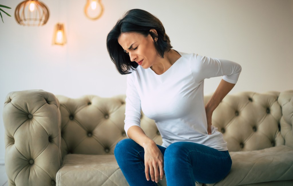
What are the symptoms of spinal stenosis?

Depending on where and how severe your spinal stenosis is, you might feel the following in your neck, back, arms, legs, hands or feet:

Spinal stenosis usually develops slowly over time. For this reason, you may not have any symptoms for a while, even if it shows up on X-rays or other imaging tests. Symptoms may come and go and affect each person differently.

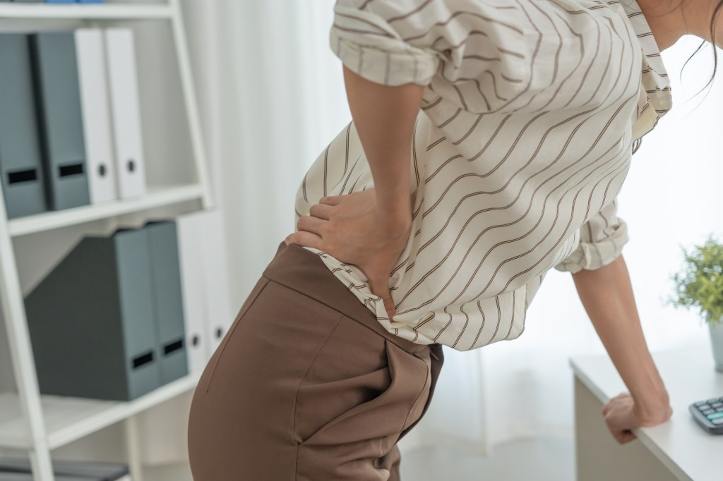
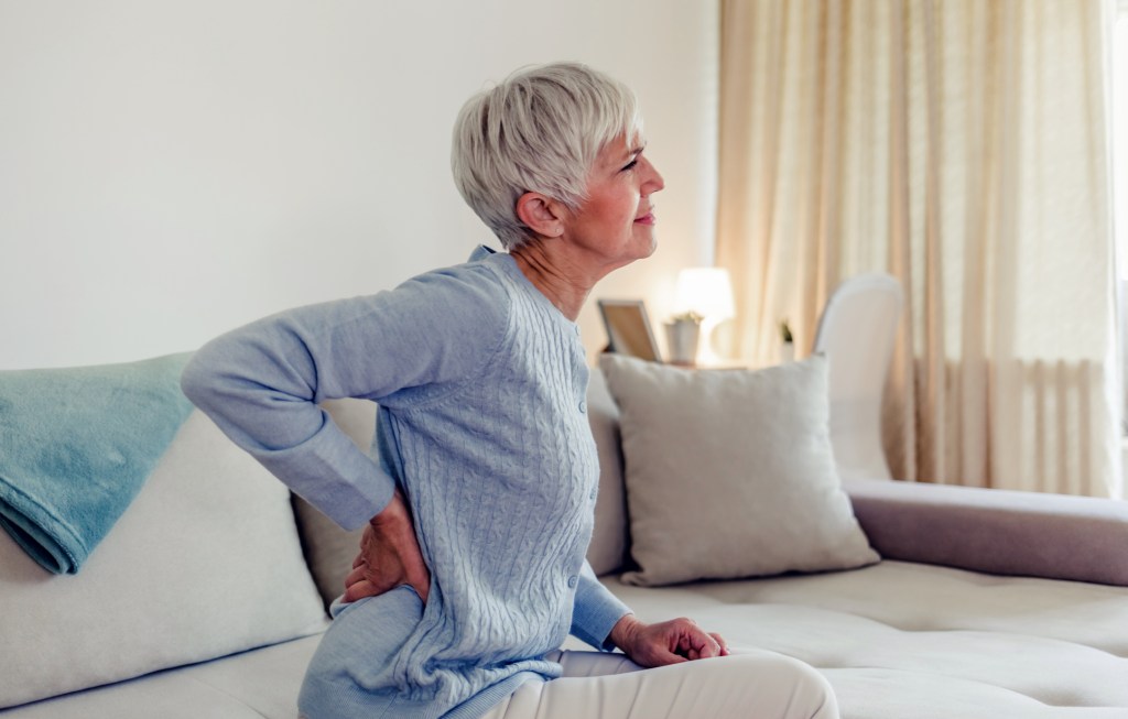
Symptoms of lumbar spinal stenosis

Symptoms of lumbar (low back) spinal stenosis include:

  • Pain in your low back.
  • Pain that begins in your buttocks and extends down your leg. It may continue into your foot.
  • A heavy feeling in your legs, which may lead to cramping in one or both legs.
  • Numbness or tingling (“pins and needles”) in your buttocks, leg or foot.
  • Pain that worsens when you stand for long periods of time, walk or walk downhill.
  • Pain that lessens when you lean forward, walk uphill or sit.

Symptoms of cervical spinal stenosis

You can feel symptoms of cervical spinal stenosis anywhere below the point of the nerve compression in your neck. Symptoms include:

  • Neck pain.
  • Numbness or tingling in your arm, hand, leg or foot.
  • Weakness or clumsiness in your arm, hand, leg or foot.
  • Balance problems.
  • Decreased function in your hands, like having issues writing or buttoning shirts.

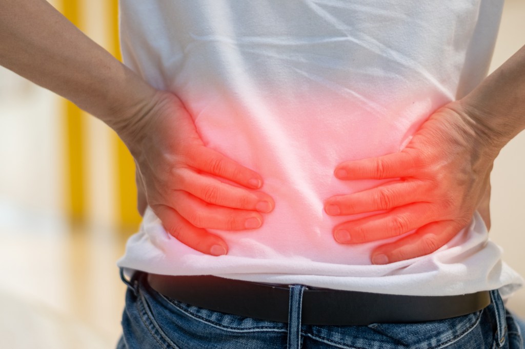
What does spinal stenosis pain feel like?

Pain from spinal stenosis can feel different from person to person. Some describe it as a dull ache or tenderness. Others describe it as an electric-like or burning sensation. The pain can come and go.

What causes spinal stenosis?

Spinal stenosis has several causes. Many different changes or injuries in your spine can cause a narrowing of your spinal canal. The causes are split into two main groups:

  • Acquired (developing after birth).
  • Congenital (from birth).

Acquired spinal stenosis is more common. It usually happens from “wear and tear” changes that naturally occur in your spine as you age. Only 9% of cases result from congenital causes.

Acquired causes of spinal stenosis

Acquired spinal stenosis means you develop it later in life (after birth) — most commonly after the age of 50. These cases usually happen from an injury or changes in your spine that occur as you age (degenerative changes).

Causes of acquired spinal stenosis include:

  • Bone overgrowth: Osteoarthritis is the “wear and tear” condition that breaks down the cartilage in your joints, including your spine. Cartilage is the protective covering of joints. As your cartilage wears away, your bones begin to rub against each other. Your body responds by growing new bone. Bone spurs, or an overgrowth of bone, commonly form. Bone spurs on your vertebrae extend into your spinal canal, narrowing the space and pinching nerves in your spine. Paget’s disease of the bone can also cause an overgrowth of bone in your spine.
  • Bulging or herniated disks: Between each vertebra is a flat, round cushioning pad (vertebral disk) that acts as a shock absorber. As you age, the disks can dry out and flatten. Cracking in the outer edge of the disks can cause the gel-like center to break through. The bulging disk then presses on the nerves near the disk.
  • Thickened ligaments: Ligaments are the fiber bands that hold your spine together. Arthritis can cause ligaments to thicken over time and bulge into your spinal canal.
  • Spinal fractures and injuries: Broken or dislocated bones in your vertebrae or near your spine can narrow your canal space. Inflammation from injuries near your spine can also cause issues.
  • Spinal cysts or tumors: Growths within your spinal cord or between your spinal cord and vertebrae can narrow your spinal canal.

Congenital causes of spinal stenosis

Congenital spinal stenosis affects babies and children. It can happen due to:

Some congenital causes of spinal stenosis include:

  • Achondroplasia: A bone growth disorder that results in dwarfism due to a genetic mutation.
  • Spinal dysraphism: When the spine, spinal cord or nerve roots don’t form properly during fetal development. Spina bifida and other neural tube defects are examples.
  • Congenital kyphosis: When your child’s spine curves outward more than it should. As a result, their upper back looks overly rounded. This happens due to an issue with fetal spine development.
  • Congenital short pedicles: When your baby is born with vertebrae pedicles (the bony “sides” of the spinal canal) that are shorter in length. This decreases their spinal canal size.
  • Osteopetrosis: A rare genetic condition that causes your child’s bones to grow abnormally and become overly dense.
  • Morquio syndrome: A rare genetic condition that affects your child’s bones, spine and other body systems.
  • Hereditary multiple exostoses (diaphyseal aclasis): A rare genetic condition that causes several small bone growths (protrusions). They can grow on your child’s vertebrae and affect their spinal canal.

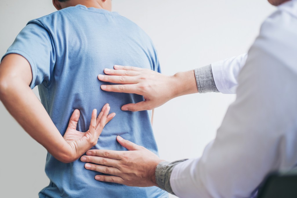
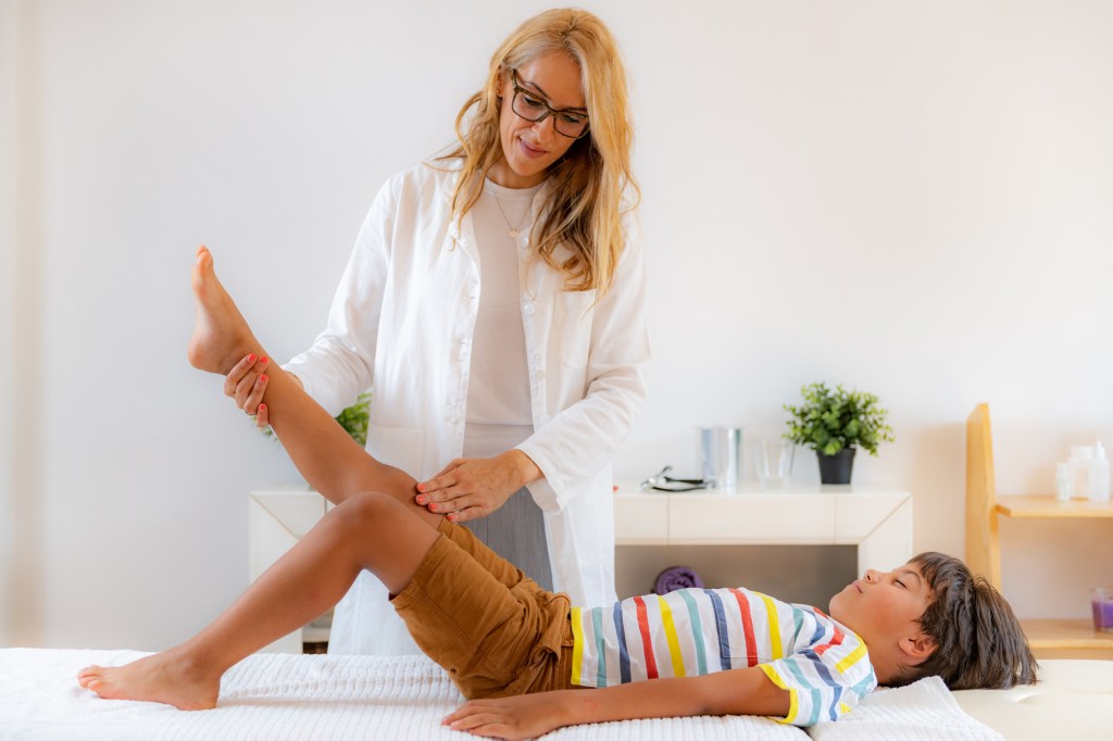
How is spinal stenosis diagnosed?

Your healthcare provider will review your medical history, ask about your symptoms and do a physical exam. Your provider may feel your spine, pressing on different areas to see if it causes pain. They’ll likely ask you to bend in different directions to see if certain spine positions bring on symptoms.

You’ll also have imaging tests so your provider can “see” your spine and determine the exact location, type and extent of the problem. These tests may include:

  • Spine X-ray: X-rays use a small amount of radiation and can show changes in bone structure. For example, they can show a loss of disk height or bone spurs.
  • MRI: Magnetic resonance imaging (MRI) uses radio waves and a powerful magnet to create cross-sectional images of your spine. MRI provides detailed images of your nerves, disks and spinal cord. It can reveal any tumors as well.
  • CT scan or CT myelogram: A computed tomography (CT) scan is a combination of X-rays that creates cross-sectional images of your spine. A CT myelogram uses a contrast dye so your provider can more clearly see your spinal cord and nerves.

What is the treatment for spinal stenosis?

There are many treatment options for spinal stenosis. What’s best for you depends on:

  • The cause.
  • The location of the issue.
  • The severity of your symptoms.

If your symptoms are mild, your healthcare provider may recommend at-home care first. If these methods don’t work and as symptoms worsen, your provider may recommend physical therapy, medications, injections and, finally, surgery.

At-home care for spinal stenosis

At-home care may include:

  • Applying heat: Heat usually is the better choice for osteoarthritis pain. Heat increases blood flow, which relaxes your muscles and relieves aching joints. Be careful when using heat — a high heat setting can burn you.
  • Applying cold: If heat isn’t easing your symptoms, try ice, like an ice pack, frozen gel pack or a frozen bag of peas. Apply the ice for 20 minutes on and 20 minutes off. Ice reduces swelling, tenderness and inflammation.
  • Exercising: Check with your healthcare provider first, but exercise can help relieve pain. It also strengthens your muscles to support your spine and improves your flexibility and balance.

Nonsurgical treatment for spinal stenosis

Nonsurgical treatments mainly help manage symptoms of spinal stenosis. They include:

  • Oral medications: Over-the-counter nonsteroidal anti-inflammatory medications (NSAIDs) can help relieve inflammation and provide pain relief from spinal stenosis. Be sure to talk with your provider to learn about the possible long-term problems of taking these medicines. Your provider may also recommend prescription medications with pain-relieving properties. These may include the antiseizure medication called gabapentin or tricyclic antidepressants, like amitriptyline. If you have muscle cramps or spasms, muscle relaxants may help.
  • Physical therapy: Physical therapists will work with you to develop a back-healthy exercise program to help you gain strength and improve your balance, flexibility and spine stability. Strengthening your back and abdominal muscles (your core) will make your spine more resilient. Physical therapists can teach you how to walk in a way that opens up your spinal canal, which can help ease pressure on your nerves.
  • Steroid injections: Getting corticosteroid injections in the space around pinched spinal nerves may help reduce inflammation, pain and irritation.

Surgery for spinal stenosis

Spinal stenosis is complex, and your spine is a delicate area. Because of this, providers consider surgery only if all other treatment options haven’t worked. Fortunately, most people who have spinal stenosis don’t need surgery.

Types of spine surgery include:

  • Laminectomy (decompression surgery): This is the most common type of surgery for spinal stenosis. It involves removing the lamina, which is a portion of your vertebra. The surgeon may also remove some ligaments and bone spurs. The procedure makes more room for your spinal cord and nerves.
  • Laminotomy: This is a partial laminectomy. The surgeon only removes a small part of the lamina — the area causing the most pressure on the nerve.
  • Laminoplasty: This surgery is just for your neck (cervical spinal stenosis). The surgeon removes part of the lamina to provide more canal space. They use metal plates and screws to create a hinged bridge across the area where they removed bone.
  • Foraminotomy: The foramen is the area in your vertebrae where the nerve roots exit. This procedure involves removing bone or tissue in this area to provide more space for the nerve roots.
  • Interspinous process spacers: This is a minimally invasive surgery for some people with lumbar spinal stenosis. The surgeon inserts spacers between the bones that extend off the back of each vertebrae called the spinous processes. The spacers help keep your vertebrae apart, creating more space for nerves.
  • Spinal fusion: Healthcare providers use spinal fusion as a last option. They only consider it if you have radiating nerve pain from spinal stenosis, your spine is not stable and other treatments haven’t helped. Spinal fusion surgery permanently joins (fuses) two vertebrae together.

Can I prevent spinal stenosis?

As most causes of spinal stenosis are normal age-related “wear and tear” conditions, you can’t totally prevent spinal stenosis. But you can take certain steps to keep your spine healthy. They may help lower your risk or slow the progression of spinal stenosis. These steps include:

  • Eating healthy foods. Be sure you’re getting enough calcium in your diet to keep your bones strong.
  • Maintaining a weight that’s healthy for you.
  • Avoiding smoking or quitting smoking. Smoking damages your arteries, which can contribute to back pain and make it difficult for any injuries to heal.
  • Practicing good posture.
  • Exercising regularly. Keeping your muscles strong, especially your back and core muscles, helps to keep your spine healthy.

What is the prognosis for spinal stenosis?

The prognosis (outlook) for spinal stenosis varies based on several factors, like:

  • Its location.
  • Its severity.
  • Your overall health.

In most cases, the prognosis for spinal stenosis is good. Many people with spinal stenosis can live full and active lives with nonsurgical treatment. But it’s important to remember that spinal stenosis affects each person differently, so not every treatment works for everyone.

What are the complications of spinal stenosis?

In severe cases, spinal stenosis can cause a loss of bladder or bowel control (incontinence). It can also cause sexual dysfunction due to nerve issues, like erectile dysfunction or anorgasmia.

It’s very rare, but extreme cases of spinal stenosis can cause partial or complete leg paralysis.

When should I see my healthcare provider about spinal stenosis?

If you notice new back pain or other symptoms, like tingling or weakness in your extremities, talk to a healthcare provider.

If you’re receiving treatment for spinal stenosis and it’s not working to help your symptoms, talk to your provider about other options.

A note from QBan Health Care Services

Back and neck pain can interrupt your daily life. The good news is that there are many treatment options for spinal stenosis. See your healthcare provider to discuss your options. They’re available to help.

SACROILIITIS

Sacroiliitis happens when something irritates the joint where your spine meets your pelvis. Arthritis is the most common cause, especially ankylosing spondylitis. Typical treatments are physical therapy and over-the-counter pain medication.

What is sacroiliitis?

Sacroiliitis is painful inflammation in the joints where your spine connects to your pelvis (your sacroiliac joints). It causes pain in your lower back, butt (your buttock muscles) or legs.

A joint is a place in your body where two bones meet. Your sacroiliac joints are the connection between your spine and pelvis. Specifically, they’re the place where the sacrum (the triangle-shaped last section of your spine) meets the ilium (the top part of your pelvis).

The sacroiliac joints are some of the biggest joints in your body, and you use them every time you move or shift your hips. Sacroiliitis happens when something irritates or damages one or both of your sacroiliac joints. This irritation leads to inflammation, which causes pain that you’ll usually feel in your low back and butt.

Visit a healthcare provider if you’re experiencing low back pain. It’s a common symptom that can be caused by a lot of issues. The sooner a provider diagnoses what’s causing your discomfort, the faster you can treat it and reduce its impact on your daily routine.

Types of sacroiliitis

A healthcare provider might refer to sacroiliitis with different names depending on how many of your sacroiliac joints are affected. Unilateral sacroiliitis is sacroiliitis that affects one of your joints. Bilateral sacroiliitis is having sacroiliitis in both joints at the same time.

How common is sacroiliitis?

It’s hard for experts to estimate how many people have sacroiliitis every year because low back pain is such a common symptom and can be caused by so many conditions. Some studies estimate that around one-quarter of people with low back pain have sacroiliitis.

What are sacroiliitis symptoms?

Pain in your lower back is the most common sacroiliitis symptom. The pain might:

  • Get worse after you’ve been sitting or standing in one position for a long time.
  • Get worse when you turn or rotate your hips.
  • Feel suddenly sharp and stabbing. You might also feel a more constant dull ache.
  • Radiate (spread) from your low back into your butt (your buttock muscles), hips or thighs.

People with sacroiliitis often feel stiff first thing in the morning. It’s common to have some stiffness after sleeping or sitting in one position, but the stiffness sacroiliitis causes usually lasts for more than an hour every time you wake up.

What causes sacroiliitis?

Anything that causes inflammation in your joints can affect your sacroiliitis joints and cause sacroiliitis. Arthritis is the main cause of sacroiliitis, including:

  • Ankylosing spondylitis: Ankylosing spondylitis is arthritis that affects the joints in your spine. Sacroiliitis is often an early symptom of ankylosing spondylitis.
  • Psoriatic arthritis: Psoriatic arthritis is a combination of psoriasis and arthritis. It causes arthritis symptoms in your joints and psoriasis (scaly, flaky patches) on your skin.

Other health conditions that cause inflammation can cause sacroiliitis too, including:

People sometimes experience sacroiliitis during pregnancy. Hormones that cause your body to change while you’re pregnant can make your sacroiliac joints widen and rotate.

A rare bacterial infection (Staphylococcus aureus) can cause sacroiliitis if the infection attacks your sacroiliac joints.

What are complications of sacroiliitis?

If it’s not treated soon enough, the pain from sacroiliitis can affect your ability to move. Untreated pain can also disrupt your sleep and lead to mental health conditions like depression.

How is sacroiliitis diagnosed?

A healthcare provider will diagnose sacroiliitis with a physical exam and some imaging tests. They’ll examine your back, hips and legs. Tell your provider when you first noticed pain or other symptoms and if certain activities make the pain worse. Talk to your provider about other health conditions or issues you have. This can help them determine what’s causing the sacroiliitis.

Your provider might have you perform some physical movements to check your range of motion (how far you can move a part of your body without feeling pain). They might press on your sacroiliac joints or the area around them. Tell your provider if any position, movement or type of pressure makes the pain worse.

What tests will providers use to diagnose sacroiliitis?

Your provider might use imaging tests to take pictures of your sacroiliac joints, including:

Your provider might also use blood tests to rule out infections and other issues that cause inflammation in your body.

How is sacroiliitis treated?

Physical therapy is the most common treatment for sacroiliitis. A physical therapist will give you stretches and exercises to strengthen the muscles around your sacroiliac joints. This will take pressure off your joints and help keep them more stable. Doing exercises for sacroiliitis will also help you increase your range of motion in your sacroiliac joints.

Your healthcare provider might also suggest you manage pain with medications, including:

  • NSAIDs: Over-the-counter nonsteroidal anti-inflammatory drugs (NSAIDs) are medications like aspirin, ibuprofen or naproxen. Don’t take NSAIDs for more than 10 days in a row without talking to your provider.
  • Muscle relaxers: Muscle relaxers (muscle relaxants) are prescription medications that treat muscle pain by preventing your nerves from sending pain signals to your brain.
  • Corticosteroids: Corticosteroids are prescription medications that relieve pain and inflammation. Your provider might inject a corticosteroid directly into your affected joint.

It’s less common, but your provider might suggest a radiofrequency ablation (RFA) to permanently block nerves in your joint from sending pain signals to your brain.

You might need to work with a rheumatologist, a specialist who treats inflammatory diseases.

Sacroiliitis surgery

It’s rare to need surgery for sacroiliitis. Your provider might recommend surgery if physical therapy and other treatments like RFA haven’t improved your pain.

Your surgeon might perform a joint fusion — permanently fastening the joint together with surgical screws. They’ll tell you which procedure you’ll need and what to expect while you’re recovering.

Can I prevent sacroiliitis?

There’s usually no way to prevent sacroiliitis.

You can lower your chances of developing arthritis by:

  • Avoiding tobacco products.
  • Doing low-impact exercise.
  • Following a diet and exercise plan that’s healthy for you.

Is sacroiliitis permanent?

If you have inflammatory arthritis, the damage in your affected joints might be permanent. Pregnant people who experience sacroiliitis usually only have it while they’re pregnant.

Most people with sacroiliitis can treat the cause with medication and manage their symptoms with physical therapy. Talk to your provider if your symptoms come back (recur) or get worse.

When should I see my healthcare provider?

Visit a healthcare provider if you’re experiencing new or worsening pain in your low back. Talk to your provider if it feels like your pain is getting worse or your sacroiliitis treatments aren’t as effective as they used to be.

What questions should I ask my healthcare provider?

  • Do I have sacroiliitis, sacroiliac joint point or another cause of lower back pain?
  • Do I have arthritis? Which type?
  • Will I need physical therapy?
  • Which medications will I need?
  • Will I need surgery?

Is sacroiliitis the same as sacroiliac joint pain?

It’s possible to have pain in or near your sacroiliac joint without having sacroiliitis. Low back pain is extremely common, and many people feel pain near their sacroiliac joint.

The difference is sacroiliitis is joint inflammation that a healthcare provider diagnoses. Visit a provider if you’re experiencing pain that doesn’t get better on its own in a week. They’ll help you understand what’s causing the pain and how you can avoid it in the future.

A note from QBan Health Care Services

Sacroiliitis is inflammation at the joint where your spine meets your pelvis, which causes low back pain. Any pain is annoying, especially when it makes it hard to move and use your body the way you usually can. The good news is that no matter what’s causing your pain, a healthcare provider will help you find ways to manage your symptoms, like physical therapy. And while physical therapy is hard work, don’t forget to celebrate your successes and take time to give yourself credit for the progress you’re making.

DEGENERATIVE DISK DISEASE

Degenerative disk disease occurs when the cushioning in your spine begins to wear away. The condition is most common in older adults. After age 40, most people experience some spinal degeneration. The right treatment can lead to pain relief and increased mobility.

What is degenerative disk disease?

Degenerative disk disease is when your spinal disks wear down. Spinal disks are rubbery cushions between your vertebrae (bones in your spinal column). They act as shock absorbers and help you move, bend and twist comfortably. Everyone’s spinal disks degenerate over time and is a normal part of aging.

When the cushions wear away, the bones can start to rub together. This contact can cause pain and other problems, such as:

How common is intervertebral disk degeneration?

Almost everyone has some disk degeneration after age 40, even if they don’t develop symptoms. It can lead to back pain in about 5% of adults.

Who might get degenerative disk disease?

Degenerative disk disease is most common in older adults. Some factors increase your risk of developing degenerative disk disease, including:

  • Acute injuries, such as falling.
  • Obesity.
  • Biological sex, with women being more likely to experience symptoms.
  • Smoking.
  • Working a physically demanding job.

What are the symptoms of degenerative disk disease?

The most common symptoms of degenerative disk disease are neck pain and back pain. You may experience pain that:

  • Comes and goes, lasting for weeks or months at a time.
  • Leads to numbness or tingling in your arms or legs.
  • Radiates down your buttocks and lower back.
  • Worsens with sitting, bending or lifting.

What causes degenerative disk disease?

Spinal disks wear down as a normal part of aging. Especially after age 40, most people experience some disk degeneration. However, not everyone experiences pain.

You might have pain if your spinal disks:

  • Dry out: Your disks have a soft core that mostly contains water. As you get older, that core naturally loses some water. As a result, disks get thinner and don’t provide as much shock absorption as they used to.
  • Tear or crack: Minor injuries can lead to small cracks in your spinal disks. These tears are often near nerves. Tears can be painful, even when they are minor. If the outer wall of your spinal disk cracks open, your disk may bulge out of place, known as a herniated disk, which may compress a spinal nerve.

What does degenerative disk pain feel like?

Degenerative disk pain:

  • Can happen in the neck or lower back.
  • May extend into the arms and hands or into the butt and legs.
  • Can be mild, moderate or severe.
  • May start and stop.
  • Can get worse after certain activities such as bending, twisting or lifting.
  • Can get worse over time.

How is degenerative disk disease diagnosed?

To diagnose degenerative disk disease, your healthcare provider may start by asking you about your symptoms. Questions may include:

  • When does the pain start?
  • Where do you feel pain?
  • What activities cause the most pain?
  • What activities decrease the pain?
  • Did you have an injury or accident that led to pain?
  • Do you have other symptoms, such as tingling or numbness?
  • How far can you walk?

Your healthcare provider may use imaging scans such as X-ray, CT or MRI. These tests can show your healthcare provider the state and alignment of your disks. Your provider may also conduct a physical exam to check your:

  • Nerve function: Your provider may use a reflex hammer to check your reactions. Poor or no reaction could mean you have damaged or compressed nerves.
  • Pain levels: Your provider may touch or press on specific areas of your back to measure your pain levels.
  • Strength: Muscle weakness or shrinking (atrophy) could mean you have nerve damage or degenerated disks.

How is degenerative disk disease treated?

Usually, your healthcare provider will recommend noninvasive treatment options first. Your treatment may include:

  • Physical therapy: Participating in strengthening and stretching exercises with a trained healthcare provider.
  • Medications: Taking nonsteroidal anti-inflammatory drugs (NSAIDs), muscle relaxers or steroids.
  • Steroid injections: Injecting medicine near your spinal nerves, disk or joints to reduce inflammation and pain.
  • Radiofrequency neurotomy: Using electric currents to burn sensory nerves and prevent pain signals from reaching your brain.

Can I treat degenerative disk disease at home?

Some people find pain relief through at-home remedies. At-home treatments may decrease pain for a short time. But they are not a long-term treatment for severely degenerated disks. You may try:

  • Exercise: Low-impact activity such as walking or swimming can strengthen back muscles and relieve some pain.
  • Hot and cold therapy: Alternating ice packs and heating pads every 10 to 15 minutes up to three to four times per day may reduce soreness and inflammation.
  • Stretching: Gentle yoga and stretching throughout the day may improve posture and relieve tension.

Do I need surgery for degenerative disk disease?

Many patients do not need surgery for degenerative disk disease. But if you have tried multiple nonsurgical treatments and have persistent pain and/or weakness, surgery may be a good option.

Or your surgeon may use one of a few types of spinal decompression surgery:

  • Diskectomy: Removing part of a spinal disk to relieve pressure on your nerves.
  • Foraminotomy: Expanding the opening for your nerve roots by removing tissue and bone.
  • Laminectomy: Taking out a small portion of bone from your lower spine (lamina).
  • Osteophyte removal: Removing bone spurs (osteophytes).
  • Spinal fusion: During this procedure, your surgeon connects two or more vertebrae to improve stability.

How can I prevent degenerative disk disease?

You can prevent or slow the progression of spinal degeneration through lifestyle changes. Some of these include:

  • Achieving and maintaining a healthy body weight.
  • Avoiding or quitting smoking.
  • Exercising regularly to increase strength and flexibility.

What is the outlook for people with degenerative disk disease?

Many people use nonsurgical and at-home treatments to manage pain long-term. If you have mild to moderate back pain, you will need to continue treatment to keep the pain at bay.

Most people who have surgery for degenerative disk disease experience long-term pain relief. Even after surgery, you need to continue exercising and stretching to keep your back strong and healthy.

Does degenerative disk disease increase my risk for other conditions?

Degenerated disks can increase your risk of developing other spinal conditions. Common spine problems include:

What else should I ask my doctor?

You may want to ask your healthcare provider:

  • What is the most likely cause of degenerative disk disease?
  • How can I slow the progression of the disease?
  • What nonsurgical treatments are most likely to relieve pain?
  • What will happen if I choose not to have surgery?
  • How can I prevent pain from returning after surgery?

A note from QBan Health Care Services

Degenerative disk disease occurs when your spinal disks break down. When these disks wear out, people typically experience back pain and stiffness. You may find pain relief with nonsurgical treatments such as physical therapy and spinal injection. For some people, home remedies like hot and cold therapy can decrease pain. When pain is severe, you may benefit from spinal injections or spine surgery. A spine specialist can help you determine which treatment is best for you.

HERNIATED DISK

A herniated disk is also known as a slipped, ruptured or bulging disk. It’s one of the most common causes of neck, back and leg pain. Most of the time, herniated disks heal on their own or with simple home-care measures.

What is a herniated disk?

A herniated disk is an injury of the spine (backbone). You have a series of bones (vertebrae) in your spine, stretching from the base of your skull to your tailbone. Between your vertebrae are round cushions called disks. The disks act as buffers between your bones, allowing you to bend and move with ease. When one of these disks tears or leaks, it’s called a herniated disk.

Every year, up to 2% of people get a herniated disk. Herniated disks are a leading cause of neck and/or arm, and back and/or leg pain (sciatica). They can happen anywhere along the spine, but herniated disks most often occur in the lower back or the neck. It’s rare for a herniated disk to be in the mid-back.

Who gets herniated disks?

People ages 30 to 50 are most likely to get a herniated disk. The problem affects men twice as often as women. Other risk factors include:

  • Sitting for long periods in the same position.
  • Being overweight.
  • Lifting heavy objects.
  • Repetitive bending or twisting motions for work, sports or hobbies.
  • Smoking.

What causes a herniated disk?

Disks have soft, gel-like centers and a firmer outer layer, like a jelly doughnut. With time, the outer layer weakens and can crack. A herniated disk happens when the inner “jelly” substance pushes through the crack. The leaked material may press on nearby spinal nerves.

Several factors can contribute to a disk rupture, including:

  • Aging.
  • Excessive weight.
  • Repetitive motions.
  • Sudden strain from improper lifting or twisting.

What are the symptoms of a herniated disk?

Herniated disk symptoms vary depending on where the problem is in your spine. Symptoms worsen with movement and get better with rest.

Herniated or slipped disk in the back (herniated lumbar disk)

It’s common for a herniated disk in the lower back to cause “sciatic nerve” pain. This sharp pain usually shoots down one side of your buttocks into your leg and sometimes the foot. Other symptoms of a herniated disk in your lower back include:

  • Back pain.
  • Tingling or numbness in the legs and/or feet.
  • Muscle weakness.

Herniated or disk in the neck (herniated cervical disk)

Symptoms of a herniated disk in your neck include:

  • Pain near or between your shoulder blades.
  • Pain that travels to your shoulder, arm and sometimes your hand and fingers.
  • Neck pain, especially in the back and on the sides of your neck.
  • Pain that increases when bending or turning your neck.
  • Numbness or tingling in your arms.

How is a herniated disk diagnosed?

Your healthcare provider will do a thorough exam. During the physical, your provider will assess your pain, muscle reflexes, sensation and muscle strength. Your provider may also order tests such as:

  • Magnetic resonance imaging (MRI): The most common and accurate imaging test for a suspected herniated disk is an MRI.
  • X-rays: Getting X-rays helps rule out other causes of back or neck pain.
  • Computed tomography (CT): A CT scan show the bones of your spine. Herniated disks can move into the space around your spinal cord and nerves and press on them.
  • Myelogram: A myelogram involves an injection of dye into your spine using X-ray guidance for a CT scan. The dye can reveal a narrowing of the spinal canal (spinal stenosis) and location of your herniated disk.
  • Electromyogram (EMG): This test involves placing small needles into various muscles and evaluate the function of your nerves. An EMG helps determine which nerve a herniated disk affects.

What can I do at home to relieve herniated disk pain?

In most cases, pain from a herniated disk can go away in time. To ease pain while your disk heals, you can:

  • Rest for one to three days, if the pain is severe, but it important to avoid long periods of bed rest to prevent stiffness.
  • Take an over-the-counter pain reliever, such as ibuprofen or acetaminophen.
  • Apply heat or ice to the affected area.

When should I see a doctor?

Initially, you can treat herniated disk pain at home. But you should see your doctor if:

  • Pain interferes with daily life, like going to work.
  • Symptoms aren’t better after four to six weeks.
  • Symptoms get worse.
  • You develop loss of bladder or bowel control.
  • You notice tingling, numbness or loss of strength in your arms, hands, legs or feet.
  • You have trouble standing or walking.

What is the best slipped disk treatment?

You may need more advanced treatments if your symptoms aren’t getting better. Your healthcare provider might recommend:

  • Medication: Your provider may prescribe an anti-inflammatory pain reliever or muscle relaxant.
  • Physical therapy: A physical therapist teaches you an exercise program to help relieve pressure on your nerves. Exercise loosens tight muscles and improves circulation.
  • Spinal injections: Called an epidural or nerve block, a spinal injection is a shot of steroid medication directly into your spine. The medication reduces swelling and inflammation of the nerve from the disk herniation. This will allow your body to heal and return to activity faster.
  • Surgery: In rare cases, a large herniated disk might injure nerves to the bladder or bowel. That may require emergency surgery. For nonemergency cases, surgery is an option when other treatments fail. There are various ways to perform spinal decompression surgery, but the goal is to relieve pressure on the nerve.

Will I need to have spinal surgery?

Herniated disks get better on their own over time or with nonsurgical treatment for 9 out of 10 people. If other treatments don’t relieve your symptoms, your healthcare provider may recommend surgery. There are multiple surgical techniques for relieving pressure on the spinal cord and nerves, including:

  • Diskectomy to remove your herniated disk.
  • Laminectomy to remove part of the bone around a herniated disk and expand your spinal canal.
  • Artificial disk surgery to replace a damaged herniated disk with an artificial one.
  • Spinal fusion to directly join two or more vertebrae together to make your spine more stable.

How can I avoid getting a herniated disk?

It’s not always possible to prevent a herniated disk. But you can reduce your risk by:

  • Using proper lifting techniques. Don’t bend at the waist. Bend your knees while keeping your back straight. Use your strong leg muscles to help support the load.
  • Maintaining a healthy weight. Excess weight puts pressure on the lower back.
  • Practicing good posture. Learn how to improve your posture when you walk, sit, stand and sleep. Good posture reduces strain on your spine.
  • Stretching. It’s especially important to take stretching breaks if you often sit for long periods.
  • Avoiding wearing high-heeled shoes. This type of shoe throws your spine out of alignment.
  • Exercising regularly. Focus on workouts that strengthen your back and abdomen muscles to support your spine.
  • Stopping smoking. Smoking can weaken disks, making them vulnerable to rupture. Consider quitting smoking.

What is the outlook for people with herniated disks?

For up to 90% of people, herniated disk pain gets better on its own or with simple medical care. You’ll probably feel better within a month. If you don’t, you should see your healthcare provider. Some people need more aggressive medical measures, such as spinal injections or surgery.

Will a herniated disk get worse?

An untreated herniated disk can get worse. That’s especially true if you continue the activities that caused it — for instance, if it developed because of your work. A worsening ruptured disk may cause chronic (ongoing) pain and loss of control or sensation in the affected area. See your healthcare provider if you still have symptoms after four to six weeks of conservative care.

What should I ask my doctor?

Questions to ask your doctor include:

  • Do I need to rest? For how long?
  • How much walking or other activities should I be doing?
  • Are there any exercises that can help?
  • What pain medication should I take?
  • Will ice or heat help?
  • If considering spine injections, what injection is likely to help relieve my pain? What are the risks?
  • If considering surgery, what are my surgical options? What are the risks?

A note from QBan Health Care Services

Having a herniated disk can make you reluctant to move. But don’t head to bed — too little movement can make pain worse when you get up again because muscles stiffen. Try to stay active and follow your healthcare provider’s recommendations for stretching. Gentle movement coupled with an over-the-counter pain reliever helps most people feel better in a few weeks.

BACK STRAINS AND SPRAINS

A back strain is an injury to either a muscle or tendon, while a back sprain is the stretching or tearing of a ligament. The symptoms, causes and treatment of back strains and sprains are discussed.

Overview

The back is a complex structure of bone and muscle, supported by cartilage, tendons and ligaments, and fed by a network of blood vessels and nerves. The back—especially the lumbar, or lower back—bears much of the body’s weight during walking, running, lifting and other activities. It makes sense, then, that injuries to the lower back—such as strains and sprains—are common.

What is a strain?

A strain is an injury to either a muscle or tendon. Tendons are the tough, fibrous bands of tissue that connect muscle to bone. With a back strain, the muscles and tendons that support the spine are twisted, pulled or torn.

What is a sprain?

A sprain is the stretching or tearing of a ligament. Ligaments are the fibrous bands of tissue that connect two or more bones at a joint and prevent excessive movement of the joint.

How common are back strains and sprains?

Strains and sprains are very common injuries. Next to headaches, back problems are the most common complaint to healthcare professionals.

What causes a back strain or sprain?

Twisting or pulling a muscle or tendon can result in a strain. It can also be caused by a single instance of improper lifting or by overstressing the back muscles. A chronic (long-term) strain usually results from overuse after prolonged, repetitive movement of the muscles and tendons.

A sprain often occurs after a fall or sudden twist, or a blow to the body that forces a joint out of its normal position. All of these conditions stretch one or more ligaments beyond their normal range of movement, causing injury.

In addition, several factors can put a person at greater risk for a back strain or sprain, including:

  • Curving the lower back excessively
  • Having overweight
  • Having weak back or abdominal muscles, and/or tight hamstrings (muscles in the back of the thighs).

Playing sports that involve pushing and pulling—such as weightlifting and football—also increases the risk of a low back injury.

What are the symptoms of a back strain or sprain?

Symptoms of a strain or sprain include:

  • Pain that gets worse when you move
  • Muscle cramping or spasms (sudden uncontrollable muscle contractions)
  • Decreased function and/or range of motion of the joint (difficulty walking, bending forward or sideways, or standing straight)

In some cases, the person may feel a pop or tear at the time of the injury.

How are back sprains and strains diagnosed?

Mild strains and sprains can usually be diagnosed based on a medical history—including a review of the symptoms and how the injury occurred—and a physical examination by a healthcare provider. In cases of more severe strains and sprains, especially when there is weakness or loss of function, an X-ray may be taken to rule out a fractured (broken) or herniated (bulging) disk as the cause of the back pain.

How are back strains and sprains treated?

The treatment for strains and sprains is similar, and often takes place in two phases.

The goal of the first phase is to reduce the pain and spasm. This may involve rest, and the use of ice packs and compression (pressure), especially for the first 24 to 48 hours after the injury. An over-the-counter nonsteroidal anti-inflammatory drug, such as ibuprofen (Motrin®), may be recommended to help reduce pain and swelling.

After the first 24 to 48 hours, returning to normal activities, as tolerated, is advisable. Extended bed rest or immobility (nonmovement) simply prolongs symptoms and delays recovery.

Most people with lumbar strain/sprain symptoms improve in about 2 weeks. If symptoms continue for more than 2 weeks, additional treatment may be required.

What complications are associated with back strains and sprains?

The most common complication of a back strain or sprain is a reduction in activity, which can lead to weight gain, loss of bone density, and loss of muscle strength and flexibility in other areas of the body.

How can back sprains and strains be prevented?

It is not possible to prevent all back injuries, but you can take some steps to help lower the risk of a sprain or strain:

  • Eat a healthy, well-balanced diet to keep your bones and muscles strong.
  • Maintain a healthy weight. Excess weight puts added stress on the structures of the lower back.
  • Exercise regularly, including stretching, to keep your joints flexible and your muscles in good condition.
  • Practice safety measures to help prevent falls, such as wearing shoes that fit properly, and keeping stairs and walkways free of clutter.
  • Use good body mechanics when sitting, standing and lifting. For example, try to keep your back straight and your shoulders back. When sitting, keep your knees bent and your feet flat on the floor. Don’t over-reach, and avoid twisting movements. When lifting, bend your knees and use your strong leg muscles to help balance the load.
  • Stop smoking. Nicotine interferes with blood flow to the muscles.

What is the prognosis (outlook) for people with back strains and sprains?

Most people with back strains and sprains have a full recovery with treatment within 2 weeks.

When should I contact my healthcare provider about a back strain or sprain?

Call your healthcare provider if:

You have severe pain and cannot walk more than a few steps.

You have numbness in the area of injury or down your leg.

You have injured your lower back several times before.

You have a lump or area with an unusual shape.

You have pain that interferes with sleep.

You have obvious weakness in an extremity (hands or feet) after an injury.

A note from QBan Health Care Services

Strains and sprains are the most common causes of back pain. You can injure muscles, tendons or ligaments by lifting something too heavy or not lifting safely. Some people strain their back by sneezing, coughing, twisting or bending over.  Back strains and sprains can be very frustrating and interrupt your daily life. There are many treatment options to help your back pain and get back to daily activities. See your healthcare provider to discuss your options. They’re there to help.

LOWER BACK PAIN

Lower back pain is very common. It can result from a strain (injury) to muscles or tendons in the back. Other causes include arthritis, structural problems and disk injuries. Pain often gets better with rest, physical therapy and medication. Reduce your risk of low back pain by keeping at a healthy weight and staying active.

What is lower back pain?

Low back pain can result from many different injuries, conditions or diseases — most often, an injury to muscles or tendons in the back.

Pain can range from mild to severe. In some cases, pain can make it difficult or impossible to walk, sleep, work or do everyday activities.

Usually, lower back pain gets better with rest, pain relievers and physical therapy (PT). Cortisone injections and hands-on treatments (like osteopathic or chiropractic manipulation) can relieve pain and help the healing process. Some back injuries and conditions require surgical repair.

How common is lower back pain?

Around four out of five people have lower back pain at some point in their lives. It’s one of the most common reasons people visit healthcare providers.

Some people are more likely to have lower back pain than others. Risk factors for lower back pain include:

  • Age: People over 30 have more back pain. Disks (soft, rubbery tissue that cushions the bones in the spine) wear away with age. As the disks weaken and wear down, pain and stiffness can result.
  • Weight: People who have overweight/obesity or carry extra weight are more likely to have back pain. Excess weight puts pressure on joints and disks.
  • Overall health: Weakened abdominal muscles can’t support the spine, which can lead to back strains and sprains. People who smoke, drink alcohol excessively or live a sedentary lifestyle have a higher risk of back pain.
  • Occupation and lifestyle: Jobs and activities that require heavy lifting or bending can increase the risk of a back injury.
  • Structural problems: Severe back pain can result from conditions, such as scoliosis, that change spine alignment.
  • Disease: People who have a family history of osteoarthritis, certain types of cancer and other disease have a higher risk of low back pain.
  • Mental health: Back pain can result from depression and anxiety.

What are the symptoms of lower back pain?

Symptoms of lower back pain can come on suddenly or appear gradually. Sometimes, pain occurs after a specific event, such as bending to pick something up. Other times, you may not know what caused the pain.

Pain may be sharp or dull and achy, and it may radiate to your bottom or down the back of your legs (sciatica). If you strain your back during an activity, you may hear a “pop” when it happened. Pain is often worse in certain positions (like bending over) and gets better when you lie down.

Other symptoms of lower back pain include:

  • Stiffness: It may be tough to move or straighten your back. Getting up from a seated position may take a while, and you might feel like you need to walk or stretch to loosen up. You may notice decreased range of motion.
  • Posture problems: Many people with back pain find it hard to stand up straight. You may stand “crooked” or bent, with your torso off to the side rather than aligned with your spine. Your lower back may look flat instead of curved.
  • Muscle spasms: After a strain, muscles in the lower back can spasm or contract uncontrollably. Muscle spasms can cause extreme pain and make it difficult or impossible to stand, walk or move.

What causes lower back pain?

Many injuries, conditions and diseases can cause lower back pain. They include:

  • Strains and sprains: Back strains and sprains are the most common cause of back pain. You can injure muscles, tendons or ligaments by lifting something too heavy or not lifting safely. Some people strain their back by sneezing, coughing, twisting or bending over.
  • Fractures: The bones in the spine can break during an accident, like a car crash or a fall. Certain conditions (such as spondylolysis or osteoporosis) increase the risk of fractures.
  • Disk problems: Disks cushion the vertebrae (small spinal bones). Disks can bulge from their position in the spine and press on a nerve. They can also tear (herniated disk). With age, disks can get flatter and offer less protection (degenerative disk disease).
  • Structural problems: A condition called spinal stenosis happens when the spinal column is too narrow for the spinal cord. Something pinching the spinal cord can cause severe sciatic nerve pain and lower back pain. Scoliosis (curvature of the spine) can lead to pain, stiffness and difficulty moving.
  • Arthritis: Osteoarthritis is the most common type of arthritis to cause lower back pain. Ankylosing spondylitis causes lower back pain, inflammation and stiffness in the spine.
  • Disease: Spine tumors, infections and several types of cancer can cause back pain. Other conditions can cause back pain, too. These include kidney stones and abdominal aortic aneurysm.
  • Spondylolisthesis: This condition causes the vertebrae in the spine to slip out of place. Spondylolisthesis leads to low back pain and often leg pain as well.

How is lower back pain diagnosed?

Your provider will ask about your symptoms and do a physical exam. To check for broken bones or other damage, your provider may order imaging studies. These studies help your provider see clear pictures of your vertebrae, disks, muscles, ligaments and tendons.

Your provider may order:

  • Spine X-ray, which uses radiation to produce images of bones.
  • MRI, which uses a magnet and radio waves to create pictures of bones, muscles, tendons and other soft tissues.
  • CT scan, which uses X-rays and a computer to create 3D images of bones and soft tissues.
  • Electromyography (EMG) to test nerves and muscles and check for neuropathy (nerve damage), which can cause tingling or numbness in your legs.
  • Depending on the cause of pain, your provider may also order blood tests or urine tests. Blood tests can detect genetic markers for some conditions that cause back pain (such as ankylosing spondylitis). Urine tests check for kidney stones, which cause pain in the flank (the sides of the low back).

What are the treatments for lower back pain?

Lower back pain usually gets better with rest, ice and over-the-counter pain relievers. After a few days of rest, you can start to get back to your normal activities. Staying active increases blood flow to the area and helps you heal.

Other treatments for lower back pain depend on the cause. They include:

  • Medications: Your provider may recommend nonsteroidal anti-inflammatory drugs (NSAIDs) or prescription drugs to relieve pain. Other medications relax muscles and prevent back spasms.
  • Physical therapy (PT): PT can strengthen muscles so they can support your spine. PT also improves flexibility and helps you avoid another injury.
  • Hands-on manipulation: Several “hands-on” treatments can relax tight muscles, reduce pain and improve posture and alignment. Depending on the cause of pain, you may need osteopathic manipulation or chiropractic adjustments. Massage therapy can also help with back pain relief and restore function.
  • Injections: Your provider uses a needle to inject medication into the area that’s causing pain. Steroid injections relieve pain and reduce inflammation.
  • Surgery: Some injuries and conditions need surgical repair. There are several types of surgery for low back pain, including many minimally invasive techniques.

Can I prevent lower back pain?

You can’t prevent lower back pain that results from disease or structural problems in the spine. But you can avoid injuries that cause back pain.

To reduce your risk of a back injury, you should:

  • Maintain a healthy weight: Excess weight puts pressure on vertebrae and disks.
  • Strengthen your abdominal muscles: Pilates and other exercise programs strengthen core muscles that support the spine.
  • Lift the right way: To avoid injuries, lift with your legs (not your back). Hold heavy items close to your body. Try not to twist your torso while you’re lifting.

What is the outlook for people with lower back pain?

The outlook depends on the cause of pain. Most people with back strains and sprains recover and do not have long-term health issues. But many people will have another episode within a year.

Some people have chronic back pain that doesn’t get better after several weeks. Older people with degenerative conditions such as arthritis and osteoporosis may have symptoms that get worse over time. Surgery and other treatments are effective at helping people with a range of injuries and conditions live pain-free.

When should I see my healthcare provider about lower back pain?

Lower back pain usually gets better with rest and pain relievers. Back pain that doesn’t go away may be a sign of a more serious condition.

See your provider if you have:

  • Pain that doesn’t get better after about a week of at-home care.
  • Tingling, numbness, weakness or pain in your buttocks or legs.
  • Severe pain or muscle spasms that interfere with your normal activities.
  • Fever, weight loss, bowel or bladder problems or other unexplained symptoms.

A note from QBan Health Care Services

Millions of people live with low back pain. Stiffness, pain and limited movement can have a major impact on quality of life. But you may be able to avoid lower back pain by maintaining a healthy weight and staying active. Talk to your provider if back pain doesn’t go away or if you’re unable to do the activities you enjoy. Several treatments can relieve pain, help you move better and get more out of life.

BACK PAIN

Pain in your back can keep you from working and living your normal life. There are so many possible reasons for your back pain that it’s wise to see your healthcare provider soon instead of trying to figure it out yourself. You may have something common like a muscle strain or there could be an underlying condition like kidney stones or endometriosis.

What is back pain?

Pain in your back can be an annoying ache, or it can get so bad that it’s unbearable. Back pain is the second most common reason why people visit their healthcare providers (just after colds). Many people miss work because of it. Around 80% to 90% of people in the United States will have back pain at some point in their lives.

What are the types of back pain?

Back pain is categorized in a number of ways by medical professionals. You can describe your back pain by its location: upper, middle or lower back pain that’s on the left side, center or right side. You may also define different types of pain to your healthcare provider. Is your pain mild, moderate or severe? Is the pain a broad ache or a smaller sharp stab? Also, back pain can be categorized by how long it lasts. An acute episode is one that is sudden and brief, and often related to an injury. Chronic/persistent means your back pain has lasted more than three to six months.

How common is back pain?

Back pain is very common. Daily, about 2% of the U.S. workforce is disabled by back pain, which is the most common reason cited for an inability to perform daily tasks.

Who is at risk for back pain?

The older you are, the more likely you are to experience back pain. You’re also at a higher risk if you:

  • Don’t exercise.
  • Already have some types of cancer or arthritis.
  • Are overweight.
  • Lift using your back instead of your legs.
  • Have anxiety or depression.
  • Smoke or use other tobacco products.

There are causes of local back pain (pain in your spine, muscles and other tissues in your back) and then there are causes of radiating back pain (pain from a problem in an organ that spreads to or feels like it’s in your back). Examples of both include:

Local back pain

Back pain in your spine may be caused by:

  • Ankylosing spondylitis.
  • Arthritis.
  • Degenerative spondylolisthesis.
  • Intervertebral disk degeneration.
  • Radiculopathy.
  • Sacroiliac joint dysfunction.
  • Spinal disk degeneration.
  • Spinal stenosis.
  • Spondylolisthesis.
  • Trauma/injury.
  • Tumor.

Back pain localized in your tissues may be caused by:

Radiating back pain

Radiating back pain may be caused by:

  • Abdominal aortic aneurysms.
  • Appendicitis.
  • Cancers (very rare).
  • Fibromyalgia and myofascial pain syndrome.
  • Infections (very rare).
  • Gallbladder inflammation.
  • Kidney infection and kidney stones.
  • Liver problems.
  • Pancreatitis.
  • Pelvic inflammatory diseases (sexually transmitted infections).
  • Perforating stomach ulcers.
  • Urinary tract infections.

In women or people assigned female at birth (AFAB), radiating back pain may be caused by:

In men or people assigned male at birth (AMAB), radiating back pain may be caused by:

  • Testicular injury or torsion.

How long does back pain last?

Back pain can last a day, a few weeks, months or a lifetime. The length of time depends on the cause and the treatment.

Is back pain a symptom of pregnancy?

It can be. Many people who are pregnant experience back pain.

Can back pain be a sign of cancer?

It’s extremely rare – only about 1% of the time will back pain be a sign of cancer.

How is back pain evaluated and diagnosed?

In many cases, your healthcare provider may get all the information they need from interviewing you about your symptoms, health history and lifestyle and then doing an exam. However, sometimes image tests are necessary. These may include:

  • X-rays.
  • MRI.
  • CT scans.
  • Bone scan.
  • EMGs.

Which healthcare providers evaluate and diagnose back pain?

Your primary healthcare provider is often able to determine the cause and diagnose your back pain. If needed, they’ll send you to a specialist and/or order tests. Possible specialists include:

  • A physical therapist.
  • An osteopath.
  • A chiropractor.
  • A medical spine provider who specializes in back pain.
  • An orthopaedic provider who specializes in bones and joints.

Your healthcare provider may also recommend a therapist or psychiatrist if you’re struggling to cope with your pain.

What questions might a healthcare provider ask to help diagnose back pain?

Your healthcare provider will ask if you injured yourself, how long you’ve had back pain and how severe your pain is. They need to know other medical problems you have and what medications you take. If you have family members who have had similar issues, let your provider know. They might also ask questions such as:

  • Are you able to work every day?
  • Does what you do for a living involve lifting?
  • Do any of your hobbies aggravate your back pain?
  • Do you have any other symptoms? (For example, if you have pain when you urinate in addition to your back pain then that may indicate a urinary tract infection.)
  • Where is your pain located?
  • How does the pain affect your daily activities?
  • What at-home treatments have you tried? (ice packs, heat pads, etc.)

How long will I have back pain?

This depends on the cause of your back pain. If your pain is caused by an infection, for example, it might go away after the course of antibiotic is complete. If your pain is caused by spinal degeneration, you may need treatment through your lifetime.

How is back pain treated?

The cause of your back pain determines the treatment. For your back pain you may feel better with:

  • Cold packs and/or heating pads.
  • Stretching exercises.
  • Massages.
  • Surgery.
  • Antibiotics.
  • Cortisone.
  • Traction.
  • Physical therapy.
  • Other over-the-counter and prescribed muscle relaxants, steroids and pain medications.
  • Exercise — specifically strengthening exercises.
  • Chiropractic care.
  • Acupuncture.

What can I do to help relieve the symptoms of back pain?

If you had an acute injury, use a cold pack for 20 to 30 minutes at a time for the first 48 hours or so. After that (or if there was no acute injury), you may find it helps more to alternate a cold pack and a heating pad. Keep one on the area for 20 to 30 minutes, and then switch. Take over-the-counter pain medications like ibuprofen (Advil®) or acetaminophen (Tylenol®). Get plenty of rest and fluids.

If you see your provider, make sure you take all medications prescribed, get all of your testing done and attend all of your appointments.

How should I sleep with back pain?

It may feel most comfortable to sleep on your back with a pillow under your knees to relieve pressure on your back. If this isn’t comfortable for you, try sleeping on your side with a pillow between your knees. Avoid sleeping on your stomach.

What can I do to reduce my risk of back pain?

Possible ways to reduce your risk of back pain include:

  • Learn to lift with your legs, not your back muscles.
  • Exercise.
  • Maintain a healthy weight.
  • Don’t slouch. Keep your back straight when you stand and sit.
  • Stop smoking and using tobacco products.
  • Get help for stress, anxiety and depression.
  • Wear low-heeled shoes.
  • Stretch regularly.

Can back pain go away on its own?

Back pain may go away on its own in some cases, but it’s best to get treatment, especially if you don’t know the cause.

When can I get back to my normal activities?

Talk to your healthcare provider about a timeline regarding when you can get back to daily activities. You may need to take time off work to rest, or you may be able to go as long as you follow your providers’ recommended treatments. Don’t guess about when you’ll be ready — confirm it with your provider.

Can back pain come back after it’s treated?

Yes, muscles and bones can get sore again.

When should I see my healthcare provider?

See your healthcare provider about your back pain if it’s severe or doesn’t get better after a few weeks. See them immediately if:

  • You feel weakness in your legs.
  • You feel numbness or tingling in your legs, genitals, buttocks or anus.
  • You’re losing weight (not on purpose).
  • There’s swelling in your back.
  • The pain spreads down one or both of your legs.

If your pain isn’t any better after four to seven days of treatment, you should contact your healthcare provider again. See your provider again as soon as possible if you now have back pain plus:

  • Blood or pus in your poop.
  • Fever.
  • Urine (pee) that is bloody or cloudy, if it smells bad or if peeing is painful.
  • Groin pain.
  • Vomiting.
  • Nausea.
  • Pain during or after sex.
  • Irregular periods.
  • Abdominal pain

What questions should I ask my healthcare provider about my back pain?

See your provider and get treatment soon so that you don’t have to suffer from back pain. Some of the questions you may want to ask them include:

  • What’s causing my back pain?
  • Is there a name for my type of back pain?
  • Will my pain go away on its own?
  • What’s my best treatment option?
  • What can I do at home to help treat my pain?
  • Do I need to see a specialist?
  • Can I work/go about my usual activities?
  • How can I prevent the back pain from coming back?

When is back pain an emergency?

Call 911 or go to the emergency department if:

  • The pain is sudden and severe.
  • You have pain and you can’t control your bowels or urine, or have nausea, fever or vomiting.
  • Your pain is so severe that you can’t go about your daily activities.

A note from QBan Health Care Services

Back pain can be very frustrating and interrupt your daily life. There are many treatment options to help your back pain and get back to daily activities. See your healthcare provider to discuss your options. They’re there to help.

ANKYLOSING SPONDYLITIS

Ankylosing spondylitis (AS) is a type of arthritis that affects the joints in your spine. It usually develops in your sacroiliac joints (where the bottom of your spine joins your pelvis). It causes typical arthritis symptoms like pain and stiffness, but it can also cause digestive symptoms, rashes and weight loss.

What is ankylosing spondylitis?

Ankylosing spondylitis is a type of arthritis that affects joints in your spine. Healthcare providers sometimes call it axial spondylarthritis.

Ankylosing spondylitis (AS) also affects the joints where the base of your spine meets your pelvis (your sacroiliac joints). Your sacroiliac joints are the connection between your spine and pelvis. Specifically, they’re the place where the sacrum (the triangle-shaped last section of your spine) meets the ilium (the top and back part of your pelvis).

The sacroiliac joints are some of the biggest joints in your body, and you use them every time you move or shift your hips.

It’s less common, but ankylosing spondylitis can affect other joints, including your:

Ankylosing spondylitis causes pain, stiffness and gastrointestinal (GI) symptoms. Visit a healthcare provider if you’re experiencing lower back pain, especially if it’s getting worse or making it hard to do all your usual daily activities.

What are ankylosing spondylitis symptoms?

Everyone with ankylosing spondylitis experiences a unique combination of symptoms. Lower back pain due to sacroiliitis (painful inflammation in your sacroiliac joints) is the most common AS symptom.

The pain can spread (radiate). You might experience other types of pain, including:

Other ankylosing spondylitis symptoms can include:

What causes ankylosing spondylitis?

Ankylosing spondylitis is an autoimmune disease. Autoimmune diseases happen when your immune system attacks your body instead of protecting it.

Experts aren’t certain what causes ankylosing spondylitis. Studies have found that specific genetic mutations are closely linked to having AS. Genetic mutations are changes to your DNA sequence that happen when your cells divide to make copies of themselves.

There are more than 60 mutated genes that might cause AS. One example is the human leukocyte antigen-B (HLA-B27) gene. More than 90% of white people who have AS also have a mutated HLA-B27 gene.

What are the risk factors?

Anyone can develop ankylosing spondylitis, but certain groups of people are more likely to have it, including:

  • People younger than 40 (more than 80% of people with AS are diagnosed when they’re around 30).
  • People assigned male at birth (AMAB).
  • People who have a close biological relative with AS (especially a biological parent).

People with certain health conditions are more likely to have ankylosing spondylitis, including:

What are the complications of ankylosing spondylitis?

People with ankylosing spondylitis have a higher risk of spinal fractures (broken bones in your spine).

Other complications can include:

  • Fused vertebrae (bones in your spine joining together).
  • Kyphosis (a forward curve in your spine).
  • Osteoporosis.
  • Eye and vision issues like uveitis or light sensitivity.
  • Heart issues, including aortitis, arrhythmia and cardiomyopathy.
  • Nerve damage.

How do providers diagnose ankylosing spondylitis?

A healthcare provider will diagnose ankylosing spondylitis with a physical exam. They’ll examine your body and discuss your symptoms. Tell your provider when you first noticed pain or other symptoms, including if any time of day or activity makes them worse.

You might need to visit a rheumatologist, a healthcare provider who specializes in treating arthritis and similar conditions.

What tests do healthcare providers use to diagnose ankylosing spondylitis?

There’s no one test that can confirm you have AS. Your provider might use a few tests to help diagnose it, including:

How is ankylosing spondylitis treated?

Your healthcare provider will suggest treatments to manage your symptoms and reduce how much they affect your daily routine.

Common treatments for ankylosing spondylitis include:

  • Exercise: Regular physical activity can reduce stiffness and stop AS from getting worse. Many people experience more severe pain when they’re inactive. A physical therapist can suggest specific stretches and exercises to help strengthen the muscles that support your back and spine.
  • Nonsteroidal anti-inflammatory drugs (NSAIDs): Over-the-counter (OTC) NSAIDs, including ibuprofen and naproxen ease pain and inflammation. Talk to your provider before taking NSAIDs for more than 10 days in a row
  • Biologic disease-modifying anti-rheumatic drugs (DMARDs): Biologic DMARDs are prescription medications that reduce inflammation and pain. They might also stop AS from getting worse.
  • Corticosteroids: Corticosteroids are prescription anti-inflammatory medications. You might need cortisone shots (an injection of corticosteroids directly into your affected joints).
  • Surgery: It’s rare to need surgery to treat AS. Your provider will usually only suggest it if you have severe symptoms that don’t get better after trying other treatments.

How soon will I feel better?

You should start feeling better soon after starting treatment. Your symptoms might not completely go away, but they should improve as you find treatments that work for you. Ask your provider when to expect improvements. Exercise and physical therapy are a gradual process, and medications take different amounts of time to take effect.

What can I expect if I have ankylosing spondylitis?

Ankylosing spondylitis is a chronic (long-term) condition. You should expect to manage your symptoms for a long time (maybe the rest of your life). Some people with AS have periods of remission where they have fewer, or milder, symptoms. Even if it’s been a while since you experienced symptoms, there’s always a chance they can come back.

There’s no cure for AS (or any other type of arthritis), but that doesn’t mean you have to live in pain. Talk to your provider about what you’re feeling. Tell them how much your symptoms impact your ability to do your favorite activities.

How do I take care of myself?

In addition to following your AS treatment regimen, there are some steps you can take to reduce inflammation in your body and stress on your joints:

  • Follow a diet and exercise plan that’s healthy for you.
  • Limit how much alcohol you drink.
  • Quit smoking.

When should I see my healthcare provider?

Visit your healthcare provider if you notice new symptoms, or if your symptoms get worse. Talk to your provider if it feels like your treatments aren’t managing your symptoms as well as they used to.

See a healthcare provider if you experience any of the following symptoms:

  • Chest pain.
  • Difficulty breathing.
  • Vision problems.
  • Severe back pain or other joint pain.
  • Your spine feels unusually stiff or rigid.
  • Unexplained weight loss.

What questions should I ask my healthcare provider?

Questions to ask your provider include:

  • Do I have ankylosing spondylitis or another type of arthritis?
  • Will I need genetic testing?
  • Which treatments will manage my symptoms?
  • Will I need physical therapy?

A note from QBan Health Care Services

Managing any type of arthritis can be frustrating. If you have ankylosing spondylitis, it’s a literal pain in the back. Pain, stiffness and other symptoms can take a toll on your day-to-day energy levels, but the good news is that AS is manageable.

Your healthcare provider will help you find treatments that reduce how much AS symptoms interfere with your day-to-day routine. Don’t be afraid to ask questions or talk to them about your treatments. You’re the best judge of how you feel.

JUVENILE IDIOPATHIC ARTHRITIS

Juvenile idiopathic arthritis is the most common type of arthritis in children. Symptoms include stiffness, pain and vision problems. Treatments include medications and physical and occupational therapy.

What is juvenile idiopathic arthritis?

Juvenile idiopathic arthritis (JIA) is the most common type of arthritis that affects children. JIA is a chronic (long-lasting) disease that can affect joints in any part of the body.

JIA is an autoimmune disease in which the body’s immune system mistakenly targets the synovium, the tissue that lines the inside of the joint, and the synovial fluid in the joint. This causes the synovium to make extra synovial fluid, which leads to swelling, pain and stiffness in the joint.

This process can spread to the nearby tissues, eventually damaging cartilage and bone. Other areas of the body, especially the eyes, may also be affected by the inflammation. If it is not treated, JIA can interfere with a child’s normal growth and development.

What are the types of juvenile idiopathic arthritis?

There are several types of JIA, depending on the symptoms and the number of joints involved:

  • Oligoarthritis: This type of JIA affects fewer than five joints, most often in the knee, ankle and elbow. It also can cause uveitis, an inflammation of the middle layer of the eye (the uvea). Uveitis may be undetectable without a dedicated eye exam. About half of all children with JIA have oligoarthritis; it is more common in girls than in boys. Many children will outgrow oligoarthritis by adulthood. In some children, it may spread to eventually involve more joints.
  • Polyarthritis: This type of JIA affects five or more joints, often the same joints on each side of the body. Polyartritis can also affect the neck and jaw joints and the smaller joints, such as those in the hands and feet. It is more common in girls than in boys and accounts for 20 percent of JIA patients
  • Psoriatic arthritis: This type of arthritis affects children who have arthritis and psoriasis, a rash that causes raised red patches or skin lesions covered with a silvery white buildup of dead skin cells. Psoriatic arthritis can also cause nail changes that look like dimpling or pitting.
  • Enthesitis-related arthritis: Also known as spondyloarthropathy. This type of arthritis often affects the spine, hips and entheses (the points where tendons and ligaments attach to bones), and occurs mainly in boys older than seven years. The eyes are often affected in this type of arthritis, and may become painful or red.
  • Systemic arthritis: Also called Still’s disease, this type occurs in about 10 to 20 percent of children with JIA. A systemic illness is one that can affect the entire body or many body systems. Besides arthritis, systemic JIA usually causes persistent high fever and rash, which most often appears on the trunk, arms and legs when fever spikes. It can also affect internal organs, such as the heart, liver, spleen and lymph nodes. This type of JIA affects boys and girls equally and rarely affects the eyes.

How common is juvenile idiopathic arthritis?

JIA is the most common type of arthritis in children. It affects about 1 in 1,000 children, or about 300,000 children in the United States.

What causes juvenile idiopathic arthritis?

The causes of JIA are not known. Factors that may be involved, alone or in combination, include genetics (the disease may be inherited), infection and environmental factors that influence the immune system.

What are the symptoms of juvenile idiopathic arthritis?

Symptoms vary depending on the type of JIA and may include:

  • Pain, swelling and tenderness in the joints. The joints may also feel warm.
  • Morning joint stiffness
  • Limping gait (younger children may not be able to perform motor activities that they recently learned)
  • Fever
  • Rash
  • Weight loss
  • Swollen lymph nodes
  • Fatigue or irritability
  • Eye redness, eye pain, and blurred vision

How is juvenile idiopathic arthritis diagnosed?

Because there are no tests for JIA, the doctor will try to rule out other causes of arthritis and other diseases as the cause of the symptoms.

To diagnose JIA, the doctor will take a complete medical history that includes a description of symptoms and perform a physical examination. The doctor will also examine the joints for signs of arthritis.

The doctor may also order these tests:

  • Imaging techniques such as X-rays or magnetic resonance imaging (MRI) to show the condition of the joints;
  • Laboratory tests on blood, urine, and/or joint fluid to help determine the type of arthritis. These include tests to determine the degree of inflammation and the presence of the substances antinuclear antibody (ANA) and rheumatoid factor. These tests also can help rule out other diseases — such as an infection, bone disorder, or cancer — or an injury as the cause of the symptoms.

How is juvenile idiopathic arthritis treated?

The goals of treatment are to relieve pain, reduce swelling, increase joint mobility and strength, and prevent joint damage and complications. Treatment generally includes medications and exercise.

Medications used to treat JIA include the following:

  • Nonsteroidal anti-inflammatory drugs (NSAIDs): These medicines provide pain relief and reduce swelling, but do not affect the course or prognosis of JIA. Some are available over the counter and others require a prescription. NSAIDs include ibuprofen (such as Motrin® and Advil®) and naproxen (such as Aleve®). These medicines can cause nausea and stomach upset in some people and need to be taken with food.
  • Corticosteroids (steroids): Corticosteroids are often given as an injection (shot) into a single affected joint. These medications can also be taken orally (by mouth) as a pill, especially by patients with more widespread disease. Oral corticosteroids can have serious side effects, including weakened bones, especially when used for long periods. Doctors usually try to limit using long-term steroids in children because they can interfere with a child’s normal growth.
  • Disease-modifying anti-rheumatic drugs (DMARDs): These medications work by changing, or modifying, the actual disease process in arthritis. The aim of DMARD therapy is to prevent bone and joint destruction by suppressing the immune system’s attack on the joints. Methotrexate is the DMARD most often used to treat JIA. Other medications used include sulfasalazine and leflunomide.
  • Biological modifying agents: Biological agents are medications that directly target molecules or proteins in the immune system that are responsible for causing the inflammation. They are given by subcutaneous injection or intravenously and are used to treat children with more severe arthritis that has not responded to other medications. Biological agents must be used with caution because they suppress the immune system and make children more vulnerable to infection. These drugs include etanercept, infliximab, adalimumab, abatacept, anakinra, rilonacept, and tocilizumab.

Exercise and physical and occupational therapy can help reduce pain, maintain muscle tone, improve mobility (ability to move) and prevent permanent handicaps. In some cases, splints or braces also may be used to help protect the joints as the child grows. Special accommodations with schools may be needed to adjust for children with limitations from their arthritis.

What complications are associated with juvenile idiopathic arthritis?

If it is not treated, JIA can lead to:

  • Permanent damage to joints
  • Interference with a child’s bones and growth
  • Chronic (long-term) arthritis and disability (loss of function)
  • Vision problems, including a loss of vision caused by uveitis (inflammation inside the eye)
  • Inflammation of the membranes surrounding the heart (pericarditis) or lungs (pleuritis) in systemic JIA

Can juvenile idiopathic arthritis be prevented?

At this time, because the cause of JIA is unknown, there is no method to prevent the development of JIA.

What is the prognosis (outlook) for children who have juvenile idiopathic arthritis?

JIA affects each child differently. For some, only one or two joints are affected and the disease is easy to control. For others, JIA may involve many joints and the symptoms may be more severe and may last longer.

With early detection and treatment, including physical and occupational therapy, it is possible to achieve good control of the arthritis, prevent joint damage, and allow normal or near-normal function for most children who have JIA.

A note from QBan Health Care Services

Children with JIA should live life as normal as possible. Attending school, playing sports, participating in extracurricular and family activities regularly is encouraged. Exercise does not make symptoms of arthritis worse. In fact, exercise can help prevent deconditioning, increase muscle strength and muscle endurance to support the joints.

There is no evidence that shows special diets are effective for arthritis, so children can eat a standard, well-balanced diet.

PSORIATIC ARTHRITIS

Psoriatic arthritis is a form of arthritis that’s linked to psoriasis — a chronic skin condition. The most common symptoms are joint pain and stiffness, skin rashes and changes in your fingernails and toenails. There’s no cure for psoriatic arthritis, but a healthcare provider will help you find treatments to manage your symptoms.

What is psoriatic arthritis?

Psoriatic arthritis is a type of arthritis. It usually affects people who have psoriasis or a biological family history of psoriasis.

Arthritis is a common disorder that affects your joints. It causes pain and inflammation in and around your joints. Psoriasis causes inflammation in your skin. Psoriasis rashes are patches of discolored skin covered with scales. These thick, scaly areas are called plaques. It can also make your fingernails and toenails thicken and look flaky or pitted (covered in tiny indentations).

Both arthritis and psoriasis are autoimmune diseases. An autoimmune disease is the result of your immune system accidentally attacking your body instead of protecting it. Psoriatic arthritis happens when you develop psoriasis symptoms on your skin and inflammation in your joints.

Psoriatic arthritis symptoms on your skin and in your joints usually come and go in periods called flares. A healthcare provider will suggest treatments to relieve your symptoms during a flare.

Visit a healthcare provider if you notice new symptoms on your skin or pain, swelling or stiffness in your joints.

How common is it?

Less than 1% of all people experience psoriatic arthritis. But it’s much more common in people who have psoriasis. Around 30% of people with psoriasis develop psoriatic arthritis at some point in their life.

What are the five forms of psoriatic arthritis?

A healthcare provider might classify psoriatic arthritis based on which joints it affects or on which side of your body you experience symptoms. The five forms of psoriatic arthritis include:

  • Distal interphalangeal predominant psoriatic arthritis: This affects the joints near the end of your fingers and toes (phalanges). It’s the most common type of psoriatic arthritis that affects your fingernails and toenails. Your nails might look discolored, flaky or pitted.
  • Symmetric polyarthritis: Polyarthritis affects five or more joints at the same time. Symmetric polyarthritis affects the same joints on both sides of your body. For example, both of your knees and both elbows. It’s one of the most common types of psoriatic arthritis.
  • Asymmetric oligoarticular psoriatic arthritis: This affects two to four joints on both sides of your body — one knee and one elbow, for example. It’s the other most common form of psoriatic arthritis (along with symmetric polyarthritis).
  • Spondylitis: Your spine is made of 33 bones called vertebrae (the plural form of vertebra). Spondylitis causes inflammation and other symptoms in the joints between your vertebrae. It can also cause pain in your hips and shoulders.
  • Arthritis mutilans: This causes severe symptoms in your hands and feet. The inflammation is usually severe enough to cause bone loss (osteolysis). It’s the rarest form of psoriatic arthritis. Less than 5% of all people with psoriatic arthritis have arthritis mutilans.

What are psoriatic arthritis symptoms?

Psoriatic arthritis can affect any joint in your body. Your symptoms can be mild — only a small amount of pain and stiffness with few signs of psoriasis on your skin. More severe psoriatic arthritis flares can make it hard or impossible to move and use your joints and will cause larger patches of psoriasis on your skin.

The most common psoriatic arthritis symptoms include:

  • Joint pain.
  • Stiffness.
  • Discoloration or redness near your affected joints.
  • Pain or tenderness where tendons and ligaments attach to your bones (like near your Achilles tendon).
  • Swelling in your fingers and toes (dactylitis or “sausage fingers”).
  • Psoriasis rash — silver or gray scaly patches on your skin (especially your scalp, elbows, knees and lower back).
  • Nail psoriasis symptoms like discoloration or pitting on your fingernails or toenails.
  • Fatigue.

What causes psoriatic arthritis?

Experts aren’t sure what causes psoriatic arthritis. Some studies have found that people with psoriatic arthritis have similar genetic mutations. Experts can’t say for certain if these changes in your genes are what cause psoriatic arthritis or if they change after you develop it.

Around 40% of people with psoriatic arthritis have a family member that also has it. This means it might be a hereditary condition — passed from biological parents to their children.

Tell your healthcare provider if someone in your family has psoriasis or psoriatic arthritis.

What are the risk factors?

Psoriatic arthritis can affect anyone. It usually develops in people between 30 and 50.

People with psoriatic arthritis typically develop psoriasis in their teens or early 20s and don’t develop symptoms in their joints until later in life.

What are psoriatic arthritis complications?

People with psoriatic arthritis are more likely to develop other health conditions, including:

Psoriatic arthritis symptoms can make it painful or difficult to use your joints. This can make it hard to get enough exercise. Talk to a healthcare provider about low-impact exercises that put less pressure on your joints. Yoga, walking or aqua therapy are all good ways to work out without stressing your joints.

How is psoriatic arthritis diagnosed?

A healthcare provider will diagnose psoriatic arthritis with a physical exam and some tests. Your provider will ask you about your symptoms and look at your skin and joints.

You might need to visit a rheumatologist, a provider who specializes in treating musculoskeletal conditions, autoimmune disorders and inflammatory conditions. A dermatologist — a provider who specializes in taking care of your skin — can diagnose and suggest treatments for psoriasis.

There’s no test that can diagnose psoriatic arthritis, but your provider might use tests to rule out other causes of your symptoms. They might use blood tests to check for infections. Imaging tests can take pictures of your joints to show damage or inflammation. You might need one or a few imaging tests, including:

How is psoriatic arthritis treated?

Your healthcare provider will suggest treatments to manage your symptoms. Which treatments you’ll need depends on:

  • Which joints the arthritis affects.
  • How severe your symptoms are.
  • How much the symptoms affect your ability to participate in your usual daily activities.

The most common treatments for psoriatic arthritis include:

Psoriatic arthritis surgery

It’s very rare to need surgery if you have psoriatic arthritis. If your joints are damaged or you’ve experienced bone loss, you might need an arthroplasty (joint replacement). Your provider or surgeon will tell you which type of surgery you’ll need and what to expect.

How can I prevent psoriatic arthritis?

Because experts don’t know what causes psoriatic arthritis, you can’t prevent it. You can lower your chances of developing all types of arthritis by:

  • Avoiding tobacco products.
  • Following a diet and exercise plan that’s healthy for you.
  • Doing low-impact, non-weight-bearing exercise.
  • Always wearing proper protective equipment for any activity that could damage your joints.

What can I expect if I have psoriatic arthritis?

You should expect to manage your symptoms for a long time (maybe the rest of your life). Some people experience long periods of time between flares, but there’s no cure for psoriatic arthritis.

It can be frustrating when a flare happens suddenly. Eventually, you might learn to recognize the warning signs of a flare and start managing symptoms before they become more severe. Talk to a healthcare provider if you feel like your symptoms are getting worse or your current treatments aren’t managing them well enough.

When should I see my healthcare provider?

Visit a healthcare provider if you notice new rashes or other symptoms on your skin or if you’re experiencing joint pain. Talk to your provider if your psoriatic arthritis symptoms are changing, flaring up more often or getting more severe.

What questions should I ask my doctor?

  • Do I have psoriatic arthritis or another type of arthritis?
  • Which tests will I need?
  • Which treatments will manage my symptoms best?
  • Should I see a rheumatologist and a dermatologist?
  • Will I need physical or occupational therapy?
  • What are good exercises to prevent muscle weakness?

Is psoriatic arthritis worse than rheumatoid arthritis?

Psoriatic arthritis and rheumatoid arthritis are different types of arthritis. They’re both autoimmune diseases.

Psoriatic arthritis is arthritis linked to psoriasis, a condition that affects your skin, fingernails and toenails. Rheumatoid arthritis is a type of arthritis that happens when your immune system attacks the tissue lining the joints on both sides of your body.

Neither condition is better or worse than the other. They’re both chronic (long-term) conditions you’ll need to manage for a long time. A healthcare provider will help you find treatments that manage your symptoms and reduce how much arthritis affects your life on a day-to-day basis.

How serious is psoriatic arthritis?

Psoriatic arthritis symptoms can range from mild to severe. You might never experience severe symptoms. But there’s a chance your symptoms will vary between flares, often with seemingly no cause or reason. Psoriatic arthritis is a chronic (long-term) condition. In that sense, it’s serious — you’ll have to manage symptoms for a long time, maybe for the rest of your life. But between flares, you should be able to participate in all your usual activities.

A note from QBan Health Care Services

It can be extremely frustrating to manage psoriatic arthritis. You might feel like you’re dealing with two conditions at the same time — the skin and nail symptoms of psoriasis and joint issues caused by arthritis. Your healthcare provider will help you find treatments that manage your symptoms during flares. They’ll also suggest ways you can keep your body healthy and strong to support your joints and skin.

Talk to your provider if it feels like your symptoms are changing, getting worse or flaring up more often. Listen to your body, and don’t hesitate to ask for adjustments in your treatment.