Tonsillitis is a common condition that happens when your tonsils get infected. Symptoms typically include sore throat, fever and swollen lymph nodes. Treatment depends on whether the infection is viral or bacterial, and recovery usually takes about one week.
What is tonsillitis?
Tonsillitis occurs when your tonsils become infected. Tonsils are the two small lumps of soft tissue — one on either side — at the back of your throat. You can see your tonsils in a mirror by opening your mouth and sticking out your tongue.
Your tonsils are part of your immune system, and they help trap germs that make you sick. When your tonsils become infected, they get swollen and sore, and swallowing may hurt. The medical term for tonsillitis is “tonsillopharyngitis”, but most people call it a sore throat because that’s what it feels like.
Tonsillitis is most common in children and adolescents, but it can affect people of all ages. It rarely occurs in children under the age of 3. Most people have tonsillitis at least once in their lifetimes.
Symptoms of tonsillitis
Tonsillitis symptoms usually come on suddenly. They may include:
Stomachache or vomiting (more common in younger children).
What are the first signs of tonsillitis?
A sore throat is often the first symptom of tonsillitis. If you develop a sudden sore throat, keep an eye on your tonsils to see if they get red or swollen.
Viral tonsillitis: Viruses like those that cause the common cold and the flu cause up to 70% of tonsillitis cases. Commonly, people with viral tonsillitis have milder symptoms than those with bacterial tonsillitis.
Bacterial tonsillitis (strep throat): Bacteria, like Group A Streptococcus, cause other cases of tonsillitis. A common name for bacterial tonsillitis is strep throat. People without tonsils can still get strep throat. (In this case, it affects their throat instead of their tonsils.) Generally, bacterial tonsillitis causes more severe symptoms than viral tonsillitis.
How does tonsillitis spread?
The viruses and bacteria that cause tonsillitis are highly contagious. They’re passed along by:
Kissing or sharing utensils, foods or drinks.
Coming into close contact with someone who’s sick.
Touching a contaminated surface and then touching your nose or mouth.
Inhaling tiny particles that become airborne when a sick person sneezes or coughs.
Risk factors
You have an increased risk of getting tonsillitis if you’re:
Between the ages of 5 and 15. Tonsillitis is most common in children and adolescents.
Exposed to germs frequently. Those who work or go to school in buildings with lots of other people have a higher risk of encountering the germs that cause tonsillitis. (Teachers who work closely with children are one example.)
Complications of tonsillitis
Tonsillitis can sometimes result in complications like:
To diagnose tonsillitis, your healthcare provider will:
Examine your throat for redness and swelling.
Ask about other symptoms you’ve had, like a fever, cough, runny nose, rash or stomachache. This can help them rule out other conditions.
Look in your ears and nose for other signs of infection.
Feel the sides of your neck to see if your lymph nodes are swollen and tender.
Tests that are used
After confirming a tonsillitis diagnosis, your provider will need to determine whether the infection is viral or bacterial. To do this, they may request a bacteria culture test.
During this procedure, your provider will swipe the back of your throat with a long cotton swab to gather cells and saliva. Then, they’ll check the sample to see if it tests positive for Group A Streptococcus bacteria. If your results are positive, you have strep throat. If your results are negative, you have viral tonsillitis.
How is tonsillitis treated?
Tonsillitis treatment depends on the cause. While symptoms of viral tonsillitis and bacterial tonsillitis can be similar, their treatments are different. Treatment may include:
Antibiotics, if your infection is bacterial. Your healthcare provider may prescribe antibiotics like penicillin, clindamycin or cephalosporin. It’s important to follow your healthcare provider’s instructions and take the full course of antibiotics, even if you’re feeling better after a couple of days. If you stop taking them too soon, the infection could get worse or spread to another part of your body.
Pain-relieving medications. Your provider may also recommend over-the-counter (OTC) pain relievers like ibuprofen or acetaminophen to help with your sore throat.
Tonsillectomy (tonsillitis surgery). If you have chronic or recurring (returning) tonsillitis, your healthcare provider may recommend a tonsillectomy. This is a procedure to surgically remove your tonsils.
Home remedies
In addition to your healthcare provider’s recommendations, you can relieve the symptoms of viral and bacterial tonsillitis by:
Drinking warm liquids, like tea, apple cider or broth.
Gargling with warm salt water.
Sucking on throat lozenges.
Can tonsillitis be prevented?
You can’t totally prevent tonsillitis. But you can reduce your risk by practicing good hygiene habits:
Wash your hands often, especially before touching your nose or mouth.
Avoid sharing foods, drinks or utensils with someone who’s sick.
Replace your toothbrush every three months and every time you get sick.
What can I expect if I have tonsillitis?
Most cases of viral tonsillitis clear up in a few days with fluids and plenty of rest. Antibiotics typically eliminate bacterial tonsillitis in about 10 days. Tonsillitis usually doesn’t cause any serious or lasting health problems.
How long does tonsillitis last?
In most cases, tonsillitis symptoms go away in three to four days. But if symptoms last longer, you should schedule a visit with your healthcare provider to rule out other, more serious issues.
When can I go back to work or school?
You should stay at home until your fever goes away and you can swallow comfortably again. This usually takes three to four days. If you’re unsure, ask your healthcare provider.
How do I take care of myself?
The best thing you can do is stay at home, get plenty of rest and drink lots of fluids. Following your healthcare provider’s guidance can ensure a speedy recovery.
When is tonsillitis an emergency?
You should contact your healthcare provider or an urgent care facility if you have:
A sore throat for more than four days.
A fever over 101 degrees F (38.33 degrees C).
Difficulty breathing.
Will tonsillitis go away on its own?
Viral tonsillitis typically goes away on its own in about one week. Bacterial tonsillitis takes about 10 days to run its course, but you’ll likely need antibiotics to reduce your risk of complications.
What does tonsillitis look like?
Tonsillitis usually causes visibly red and inflamed tonsils. In some cases, you might have a whitish coating on your throat or white spots on your tonsils.
Tonsillitis vs. strep: What’s the difference?
Strep throat is another common name for bacterial tonsillitis. You can get strep throat even if you don’t have your tonsils anymore.
A note from QBan Health Care Services
You know that feeling — that scratchy sensation in the back of your throat. You keep your fingers crossed, hoping it’ll go away. But when you wake up the next morning, it hurts to swallow. If this sounds like you, it could be tonsillitis. And it’s best to make an appointment with your healthcare provider. With some rest and medication, you’ll be feeling like yourself again in a few days.
Tuberculosis is a bacterial infection that is also known as TB. It can be fatal if not treated. TB most often affects your lungs, but can also affect other organs like your brain.
What is tuberculosis?
Tuberculosis is an infectious disease that can cause infection in your lungs or other tissues. It commonly affects your lungs, but it can also affect other organs like your spine, brain or kidneys. The word “tuberculosis” comes from a Latin word for “nodule” or something that sticks out.
Tuberculosis is also known as TB. Not everyone who becomes infected with TB gets sick, but if you do get sick you need to be treated.
If you’re infected with the bacterium, but don’t have symptoms, you have inactive tuberculosis or latent tuberculosis infection (also called latent TB). It may seem like TB has gone away, but it’s dormant (sleeping) inside your body.
If you’re infected, develop symptoms and are contagious, you have active tuberculosis or tuberculosis disease (TB disease).
The three stages of TB are:
Primary infection.
Latent TB infection.
Active TB disease.
How common is tuberculosis?
About 10 million people became ill with TB throughout the world, and about 1.5 million people died from the disease in 2020. TB was once the leading cause of death in the U.S. but the number of cases fell rapidly in the 1940s and 1950s after researchers found treatments.
Statistics show that there were 7,860 tuberculosis cases reported in the U.S. in 2021. The national incidence rate is 2.4 cases per 100,000 people.
Are there different kinds of tuberculosis?
In addition to active or inactive, you might hear about different kinds of TB, including the most common, pulmonary (lung) tuberculosis. But the bacterium can also affect other parts of your body besides the lungs, causing extrapulmonary tuberculosis (or TB outside of the lung). You might also hear about systemic miliary tuberculosis, which can spread throughout your body and cause:
Meningitis, an inflammation of your brain.
Sterile pyuria, or high levels of white blood cells in your urine.
Pott’s disease, also called spinal tuberculosis or tuberculosis spondylitis.
Addison’s disease, an adrenal gland condition.
Hepatitis, a liver infection.
Lymphadenitis in your neck, also called scrofula or TB lymphadenitis.
What causes tuberculosis?
TB is caused by the bacterium Mycobacterium tuberculosis. The germs are spread through the air and usually infect the lungs, but can also infect other parts of the body. Although TB is infectious, it doesn’t spread easily. You usually have to spend a lot of time in contact with someone who is contagious in order to catch it.
How is tuberculosis spread?
TB can be spread when a person with active TB disease releases germs into the air through coughing, sneezing, talking, singing or even laughing. Only people with active pulmonary infection are contagious. Most people who breathe in TB bacteria are able to fight the bacteria and stop it from growing. The bacterium becomes inactive in these individuals, causing a latent TB infection.
As many as 13 million people in the U.S. have latent TB. Although the bacteria are inactive, they still remain alive in the body and can become active later. Some people can have a latent TB infection for a lifetime, without it ever becoming active and developing into TB disease.
However, TB can become active if your immune system becomes weakened and cannot stop the bacteria from growing. This is when the latent TB infection becomes active TB. Many researchers are working on treatments to stop this from happening.
What are the signs and symptoms of tuberculosis?
People with inactive TB do not exhibit symptoms. However, they may have a positive skin reaction test or blood test.
Those with active TB can show any of the following symptoms:
What kinds of tests are used to diagnose tuberculosis?
There are two kinds of screening tests for TB: the Mantoux tuberculin skin test (TST) and the blood test, called the interferon gamma release assay (IGRA).
For the TST, a healthcare provider will inject a small amount of a substance called purified protein derivative (PPD) under the skin of your forearm. After two to three days, you must go back to the healthcare provider, who will look at the injection site.
For the IGRA, a healthcare provider will draw blood and send the sample to the lab.
Further tests to determine if an infection is active or if your lungs are infected include:
How do I know if I should get tested for tuberculosis?
You may want to get tested for TB if:
You are a resident or employee in group settings where the risk is high, such as jails, hospices, skilled nursing facilities, shelters and other healthcare facilities.
You work in a mycobacteriology laboratory.
You’ve been in contact with someone who’s known or suspected to have TB disease.
Your body’s resistance to illness is low because of a weak immune system.
You think you might already have TB disease and have symptoms.
You’re from a region or have lived in a region where TB disease is prevalent, such as Latin America, the Caribbean, Africa, Asia, Eastern Europe and Russia.
You’ve injected recreational drugs.
Your healthcare provider recommends testing.
Others who are at risk for TB include:
People with immature or impaired immune systems, such as babies and children.
People with kidney disease, diabetes, or other chronic (long-term) illness.
People who have received organ transplants.
People being treated with chemotherapy for cancer or other types of treatments for immune system disorders.
The incidence rates for minority groups in the U.S. are higher than the incidence rates for whites.
How is tuberculosis treated?
TB infection and disease is treated with these drugs:
Isoniazid (Hyzyd®).
Rifampin (Rifadin®).
Ethambutol (Myambutol®).
Pyrazinamide (Zinamide®).
Rifapentine (Priftin®).
You must take all of the medication your provider prescribes, or not all of the bacteria will be killed. You will have to take these medications for as long as you’re told — sometimes up to nine months.
Some forms of TB have become resistant to medications. It’s very important and likely that your provider will use more than one drug to treat TB. It’s very important to finish your entire prescription.
Complications/side effects of treatment
Some people have side effects from the drugs used to treat TB that may include:
Talk to your provider about any side effects because some might mean that you’re experiencing liver damage.
How soon after starting treatment for active TB will I feel better?
It will probably take weeks before you start having more energy and fewer days with symptoms. However, it will take longer than that to complete your treatment. You’ll need to take your medications for at least six to nine months.
Can tuberculosis be cured?
Yes, TB is curable.
What can you do to prevent spreading tuberculosis?
You usually have to be in contact with someone with active TB for a long time before becoming infected. It helps to follow infection prevention guidelines like:
Washing your hands thoroughly and often.
Coughing into your elbow or covering your mouth when you cough.
Avoiding close contact with other people.
Making sure you take all of your medication correctly.
Not returning to work or school until you’ve been cleared by your healthcare provider.
In the hospital, the most important measures to stop the spread of TB are having proper ventilation and using the correct types of personal protective equipment.
Is there a vaccine to prevent tuberculosis?
Some countries (but not the U.S.) use a TB vaccine called Bacillus Calmette-Guerin (BCG). The vaccine is mostly given to children in countries with high rates of TB to prevent meningitis and a serious form of TB called miliary tuberculosis. The vaccine may make skin tests for TB less accurate.
What is the outlook (prognosis) for someone with tuberculosis?
If you have tuberculosis and you’re treated, your outlook is good if you’ve followed directions and taken your medications for as long as you should and in the way you were told.
When should I see my healthcare provider?
If you’ve been exposed to TB, you should talk to your healthcare provider right away. They can help you make a decision about getting tested. That decision is more important if you’ve developed any symptoms of illness that could mean you’re contagious. Remember, even though tuberculosis can be treated, it can also be fatal if it’s not treated.
A note from QBan Health Care Services
Tuberculosis is an infection that is spread through the air. Even though it can be treated, it’s still responsible for many deaths around the world. Make sure you contact your healthcare provider if you think you’ve been exposed or have symptoms of TB. Also, make sure to follow instructions if you’re treated for TB. Ask your healthcare provider if you have any questions.
Whooping cough (pertussis) is a very contagious respiratory infection that causes a distinctive “whooping” cough. The condition can cause bouts of repeated, violent coughing that may come and go and last for weeks or months. Pertussis can cause severe, life-threatening illness, especially in babies. The pertussis vaccine can help prevent it.
What is whooping cough?
Whooping cough, also called pertussis, is a very contagious upper respiratory infection. It usually gives you lengthy and repeated bouts of coughing. These coughing episodes can continue for weeks or even months after you first develop symptoms of the illness.
Instead of coughing spells, babies with pertussis may have breathing difficulties. This includes conditions such as apnea, when there are pauses in their breathing. Whooping cough can cause serious, life-threatening complications in babies. About one-third of all infants (babies younger than 1 year old) who get whooping cough need treatment at a hospital.
Prolonged coughing causes air to be expelled from your lungs. When you gasp for air quickly and deeply after a coughing fit, a whooping cough sound might accompany the inhalation of air. This sound is a loud, high-pitched “whooping” noise. That’s where pertussis gets its name. However, someone may still have the infection without making the noise.
Who does whooping cough affect?
Pertussis can affect anyone, but it most often occurs in babies, children and adolescents. Babies are especially vulnerable to infection because they can’t receive the pertussis vaccine until they’re at least 2 months old. They can catch whooping cough from their parents, adult caregivers or other children.
Can adults get whooping cough?
Yes. But whooping cough in adults is generally milder than in babies and children. This is especially true for adults who’ve received the whooping cough vaccine. The infection may seem more like the common cold. The “whoop” may not be there in people with milder illnesses.
However, adults can develop serious cases of whooping cough, especially if they haven’t received the pertussis vaccine. They may have long-lasting coughing fits that keep them awake all night. People who’ve experienced these coughing bouts say it’s the worst cough of their lives. It can also cause major interruptions in your daily life and serious complications.
How common is pertussis?
Before the development of the pertussis vaccine, there were hundreds of thousands of cases of whooping cough each year in the U.S.
Today, case numbers have dropped significantly. According to the Centers for Disease Control and Prevention (CDC), there were about 1,600 reported cases of pertussis in the U.S. in 2021.
Every few years, outbreaks occur, and there are peaks in reported cases. In addition, whooping cough continues to be a global endemic. According to the World Health Organization (WHO), there were more than 151,000 cases of pertussis worldwide in 2018.
What are the symptoms of whooping cough?
Early pertussis symptoms may resemble those of the common cold. These symptoms may persist for one to two weeks and may include:
Slight fever.
Mild or occasional coughing.
Runny nose.
A pause in breathing in babies (apnea).
Whooping cough symptoms after the first or second week have passed usually include:
Prolonged, repeated or violent coughing episodes (paroxysms) that recur intermittently for up to 10 weeks or more.
Whooping sound when inhaling after the coughing stops.
Vomiting.
Exhaustion due to prolonged coughing.
Symptoms of pertussis begin to lessen after four weeks, although bouts of coughing can recur for months after symptoms start.
What causes whooping cough?
A type of bacteria called Bordetella pertussis causes whooping cough. The condition starts when the bacteria enter your respiratory system. They attach to the tiny, hair-like extensions (cilia) on the lining of your respiratory tract. Then, they release poisons (toxins), which damage your cilia and cause your airways to swell. This swelling causes the secretions of your mucus to increase, which causes severe coughing.
Is whooping cough contagious?
Yes. When you cough or sneeze, you can spread tiny respiratory droplets containing the bacteria into the air. People around you may inhale these bacteria-containing droplets and get infected. You may be contagious even before symptoms appear and remain contagious for up to two weeks after coughing starts.
How is whooping cough diagnosed?
A healthcare provider will perform a physical examination and ask questions about your symptoms. They may use a cotton swab to take a sample of the mucus inside your nose. They can also collect a sample by filling a syringe with saline fluid and flushing it through your nose and the back of your throat.
The provider will send the samples to a lab where a technician will analyze them for the presence of Bordetella bacteria. The provider may also request blood tests to confirm the presence of the bacteria.
What is the treatment for whooping cough?
Whooping cough treatment should begin as soon as possible after diagnosis. A provider will prescribe antibiotics to help prevent the spread of the disease. But antibiotics can’t prevent or treat your cough. Cough syrups and other medicines can’t relieve your coughing spells, so you’ll need to use other forms of home treatment to manage your symptoms.
If you’ve been in close contact with someone infected with pertussis, you should start treating whooping cough within three weeks of exposure. A provider will likely recommend antibiotics as well.
If your baby develops the condition, they may need pertussis treatment in the hospital. Whooping cough can cause life-threatening complications, such as pneumonia or breathing problems. To treat the infection and prevent complications, a healthcare provider will:
Keep your baby’s breathing passages clear. They may have to suction out mucus.
Monitor your baby’s breathing. They’ll give them oxygen, if necessary.
Prevent or treat dehydration. Your baby may need an IV to receive fluids.
How can I prevent whooping cough?
Getting the whooping cough vaccine is the best way to prevent it getting whooping cough. Healthcare providers recommend that all children receive the diphtheria-tetanus-pertussis (DTaP) vaccine. This is a combination vaccine that also protects them against diphtheria and tetanus. It’s safe and effective.
Children should receive five injections of the DTaP vaccine, according to the following recommended vaccination schedule:
First dose: 2 months old.
Second dose: 4 months old.
Third dose: 6 months old.
Fourth dose: Between 15 and 18 months old.
Fifth dose: Between 4 and 6 years old.
Healthcare providers recommend a booster vaccine for adults because they’re the most likely source of pertussis infection in infants. It’s important that all adults caring for infants receive a booster, as adults’ immunity to pertussis wanes as they get older. Adults who have a whooping cough infection may be told they have bronchitis or a sinus infection, and may unknowingly infect susceptible infants who are at risk for more serious complications.
Adults between the ages of 19 and 64 should receive a one-time booster pertussis vaccine called the Tdap vaccine.
Adults older than 64 years old should also receive a booster vaccine if they’ll come into close contact with babies younger than 12 months old. Pregnant people should receive a Tdap injection during the third trimester (between the 27th and 36th week) of their pregnancies. They must have a Tdap injection during each pregnancy.
The vaccine helps provide babies with short-term protection against whooping cough. It may also protect them against serious complications associated with the infection. If you’re pregnant, you should also make sure family members and caregivers take pertussis precautions by getting vaccinated.
What can I expect if I have whooping cough?
Pertussis may initially look like the common cold. But eventually, the telltale sign of the condition — the whooping cough sound — may develop. With prompt diagnosis, you can start on antibiotics that can help prevent the spread of the infection. But antibiotics won’t treat the cough. Your coughing episodes may last for weeks or even months. Whooping cough is more serious in babies and children, and serious complications can develop.
What are the possible complications of whooping cough?
Whooping cough can lead to serious, life-threatening complications, especially in babies younger than 1 year old. Approximately one-third of infants will need care in the hospital. Of those babies, about:
2 in 3 (68%) will develop pauses in their breathing called apnea.
1 in 5 (22%) will develop pneumonia.
1 in 50 (2%) will have violent, uncontrollable episodes of shaking (convulsions).
1 in 150 (0.6%) will develop a brain disease called encephalopathy.
1 in 100 (1%) will die.
Adults can also develop complications, but they’re usually less severe. If you have a persistent, violent cough, you may:
Pass out.
Break (fracture) a rib.
Lose your bladder control.
Lose weight unintentionally.
How do I take care of myself?
If you have whooping cough, there are several steps you can take to help manage your symptoms at home:
Get plenty of rest.
Drink lots of fluids to prevent dehydration.
Use a cool-mist humidifier to soothe your lungs and loosen mucus in your respiratory tract.
Take the antibiotics your provider prescribed as directed.
Don’t take cough medicine unless recommended by your provider.
Keep your home free from irritants such as smoke and dust that may trigger coughing fits.
When should I see my healthcare provider?
If you develop any signs of dehydration, you should seek advice from your healthcare provider immediately. Symptoms of dehydration may include:
Dry mouth.
Excessive thirst.
Fatigue.
Peeing less (decreased urination).
Muscle weakness.
Headache.
Dizziness or lightheadedness.
A note from QBan Health Care Services
Whooping cough (pertussis) is a respiratory infection that can cause severe, prolonged coughing episodes or breathing difficulties. It can cause life-threatening illnesses, especially in babies. If you or your child develop a loud, “whooping” cough or you notice your baby is having trouble breathing, seek treatment right away. And if you haven’t already, get the pertussis vaccine to prevent infection.
Some days (most days, honestly) coffee can feel like the glue that’s holding it all together. It turns you from zombie to human in the morning. It gives you a nice afternoon pick-me-up, and keeps you moving all those hours in between.
But — deep breath — is coffee good for you?
You can exhale.
Coffee, it turns out, packs some surprising health benefits.
There aren’t a lot of downsides to drinking moderate amounts of coffee — and in fact, it can have positive effects on your health. You don’t need another reason to pour yourself a fresh cup. But just in case, keep reading. (Go ahead and refresh your mug first if you’d like. We can wait a minute…)
Is coffee good for you?
The various ingredients in coffee add up to a drink that’s greater than the sum of its parts. And while how much coffee you drink, and how you take it, will make a difference in its health benefits, there are several ways your cup of joe can do your body good.
Caffeine’s positive side
Coffee gets its kick from caffeine, a natural stimulant that makes you feel more energetic.
Caffeine can get a bad rap, but it turns out the caffeine in coffee does more than just wake you up.
It acts on your brain to improve memory, mood, reaction times and mental function. One study even says caffeine can improve endurance and performance during exercise.
Nutrients
Coffee contains about a thousand different botanical compounds. Scientists haven’t studied all of them well, but the news so far gets two thumbs up.
Coffee comes from beans, after all. And dietitians love beans.
Coffee is a source of nutrients, including B vitamins, potassium and riboflavin.
Phenolic compounds
Coffee beans are also rich in antioxidants, which are compounds that protect cells against damage.
But there’s more to it than that. The latest research is showing that it’s not the antioxidants per se that make coffee, in moderation, beneficial. It’s actually the phenolic compounds.
Phenolic compounds, or phenols, are substances found in plant foods that play a key role in your body’s defense systems, protecting it from oxidative stress, as well as inflammation.
Coffee is made from beans, a plant food. Research is showing that the phenolic components in coffee provide health-promoting effects similar to those in vegetables or fruits.
Lowered risk for diabetes
Multiple studies have shown that regular coffee consumption lowers the odds of developing Type 2 diabetes. That’s true for decaf, as well as the high-octane variety.
Prevent neurologic disease
Regular daily caffeine intake — like the kind you get from your daily cuppa — is linked to a lower risk of developing Alzheimer’s disease, as well as Parkinson’s disease.
Liver benefits
Coffee has been shown to be a positive for your liver health. It can help protect against liver cirrhosis in people at risk of the disease, such as those with alcohol use disorder or fatty liver disease.
Lower cancer risk
Researchers have found that coffee drinkers have a lower risk of liver cancer and colorectal cancer — two of the leading causes of cancer deaths in the world.
Ward off depression
That pick-me-up you get from a foamy cappuccino may not be a figment of your imagination. Several studies have found that the more coffee a person drinks, the lower their risk of depression.
Are there risks associated with coffee?
Coffee may be a healthy little bean, but it isn’t an all-out magical one. To get more pros and fewer cons, here are some guidelines.
Don’t go overboard
Excessive caffeine can cause dehydration. But it’s worth noting that a moderate amount of coffee isn’t dehydrating, contrary to popular opinion.
At modest levels of intake, coffee is a net positive in terms of hydration, meaning it still contributes to your fluid needs.
Some people may find caffeine makes them jittery or anxious. And too much caffeine can also interfere with a good night’s sleep, especially if you drink it late in the day.
Coffee can also impact blood pressure. Whether it will depends on the amount you drink and on your genetic makeup. People are genetically predisposed to be slow or fast metabolizers of caffeine, and slow metabolizers may experience negative effects, such as elevated blood pressure, even at low levels of intake.
For the general population, experts recommend sticking to less than 400 milligrams of caffeine per day. An 8-ounce cup of coffee typically has 80 to 100 mg of caffeine. So, aim for about three to four cups a day, max.
Caffeine in coffee can stay in your system for several hours after your last sip. So a late-afternoon latte or post-dinner café au lait may leave you tossing and turning at night. And sleep deprivation is nothing to mess with. To play it safe, stick to decaf in the evening.
Choose wisely
If you enjoy coffee drinks, the safest way to go is to ask for sugar-free syrups and no whip with nonfat milk.
Coffee may be healthy, but what you add to it often isn’t. Keep an eye on the sugar and saturated fat, especially if you’re drinking multiple mugs a day.
Here are some suggestions to get the most health benefits from your coffee, without the additions:
Aim for natural sweeteners
There are big differences in the health implications for various sweeteners.
If you prefer artificial sweeteners, like sucralose (Splenda®) saccharin (Sweet’N Low®) and aspartame (Equal®, NutraSweet®), slowly wean yourself from them and opt for more natural sugars.
Over time, research shows that artificial sweeteners can train your brain to want very sweet things. So, it can be a tough transition to make, but it can be a major win for your health to let go of artificial sweeteners.
If you take your coffee with regular sugar but want to avoid unnecessary calories, try switching to a small amount of honey, agave or even Stevia™. Stevia comes from a leaf and is a slightly more natural source than a typical artificial sweetener.
Spice it up
Spices aren’t just a great way to add flavor to coffee, but they can also provide surprising health benefits.
Cardamom is a good source of vitamin C, calcium, magnesium, potassium and zinc. It’s also a good source of dietary fiber, iron and manganese.
Cocoa powder is a good source of protein, potassium and zinc, as well as a good source of dietary fiber, iron, magnesium, phosphorus, copper and manganese. But it’s also high in saturated fat, so use it sparingly. And go for the unsweetened kind.
Be careful with whole milk
You might wonder if it’s really a big deal to use whole milk in your coffee if it’s only a few tablespoons?
Yes, it undoubtedly matters. Those are empty, unnecessary calories that add up.
Let’s do some math. If you used two tablespoons of whole milk in four cups of coffee per day, that’s the equivalent of a half a cup of whole milk, which is 75 calories. Over a year, that would mean you’re taking in an extra 27,000 calories. (For perspective, that’s about how many calories most people should consume over the course of two weeks.)
If you want a healthier alternative for whitening up your coffee, try 1% or nonfat cow’s milk. You can also go with low-fat soy, almond or rice milk, which are all good alternatives, but are lower in protein than cow’s milk.
A Note from QBan Health Care Services
While coffee is a pleasurable part of your lifestyle, there are other factors that make a bigger impact on your health such as eating a balanced diet, exercising and maintaining a healthy weight. But drinking coffee is a delightful addition to those key health factors.
So, yes, go on and enjoy that morning coffee with the confidence that you’re doing a good thing for your body. Just don’t go overboard.
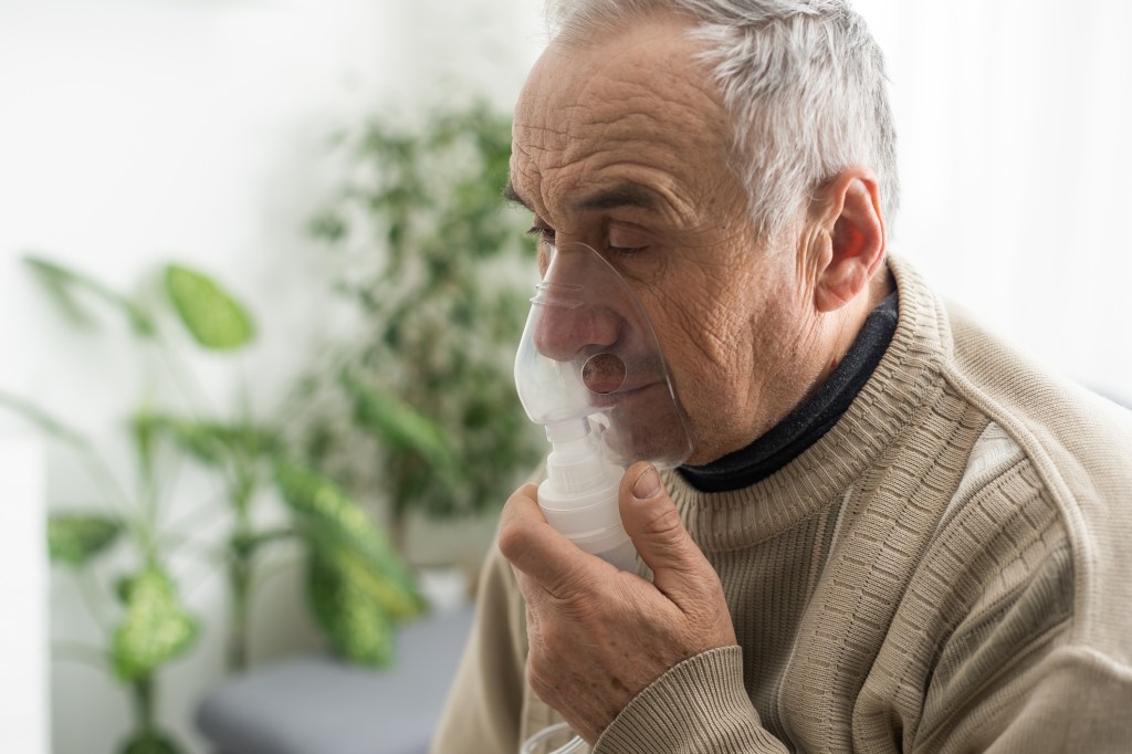
Pneumonia is inflammation and fluid in your lungs caused by a bacterial, viral or fungal infection. It makes it difficult to breathe and can cause a fever and cough with yellow, green or bloody mucus. The flu, COVID-19 and pneumococcal disease are common causes of pneumonia. Treatment depends on the cause and severity of pneumonia.
What is pneumonia?
Pneumonia is an infection in your lungs caused by bacteria, viruses or fungi. Pneumonia causes your lung tissue to swell (inflammation) and can cause fluid or pus in your lungs. Bacterial pneumonia is usually more severe than viral pneumonia, which often resolves on its own.
Pneumonia can affect one or both lungs. Pneumonia in both of your lungs is called bilateral or double pneumonia.
What’s the difference between viral and bacterial pneumonia?
While all pneumonia is inflammation caused by an infection in your lungs, you may have different symptoms depending on whether the root cause is a virus, bacteria or fungi.
Bacterial pneumonia tends to be more common and more severe than viral pneumonia. It’s more likely to require a hospital stay. Providers treat bacterial pneumonia with antibiotics. Viral pneumonia causes flu-like symptoms and is more likely to resolve on its own. You usually don’t need specific treatment for viral pneumonia.
What are the types of pneumonia?
We categorize pneumonia by which pathogen (virus, bacteria or fungi) caused it and how you got it — community-acquired, hospital-acquired or ventilator-associated pneumonia.
Community-acquired pneumonia (CAP)
When you get pneumonia outside of a healthcare facility, it’s called community-acquired pneumonia. Causes include:
Bacteria: Infection with Streptococcus pneumoniae bacteria, also called pneumococcal disease, is the most common cause of CAP. Pneumococcal disease can also cause ear infections, sinus infections and meningitis. Mycoplasma pneumoniae bacteria causes atypical pneumonia, which usually has milder symptoms. Other bacteria that cause CAP include Haemophilus influenza, Chlamydia pneumoniae and Legionella (Legionnaires’ disease).
Viruses: Viruses that cause the common cold, the flu (influenza), COVID-19 and respiratory syncytial virus (RSV) can sometimes lead to pneumonia.
Fungi (molds): Fungi, like Cryptococcus, Pneumocystis jirovecii and Coccidioides, are uncommon causes of pneumonia. People with compromised immune systems are most at risk of getting pneumonia from a fungus.
Protozoa: Rarely, protozoa like Toxoplasma cause pneumonia.
Hospital-acquired pneumonia (HAP)
You can get hospital-acquired pneumonia (HAP) while in a hospital or healthcare facility for another illness or procedure. HAP is usually more serious than community-acquired pneumonia because it’s often caused by antibiotic-resistant bacteria, like methicillin-resistant Staphylococcus aureus (MRSA). This means HAP can make you sicker and be harder to treat.
Healthcare-associated pneumonia (HCAP)
You can get HCAP while in a long-term care facility (such as a nursing home) or outpatient, extended-stay clinics. Like hospital-acquired pneumonia, it’s usually caused by antibiotic-resistant bacteria.
Ventilator-associated pneumonia (VAP)
If you need to be on a respirator or breathing machine to help you breathe in the hospital (usually in the ICU), you’re at risk for ventilator-associated pneumonia (VAP). The same types of bacteria as community-acquired pneumonia, as well as the drug-resistant kinds that cause hospital-acquired pneumonia, cause VAP.
Aspiration pneumonia
Aspiration is when solid food, liquids, spit or vomit go down your trachea (windpipe) and into your lungs. If you can’t cough these up, your lungs can get infected.
How can I tell if I have pneumonia versus the common cold or the flu?
It can be difficult to tell the difference between the symptoms of a cold, the flu and pneumonia, and only a healthcare provider can diagnose you. As pneumonia can be life-threatening, it’s important to seek medical attention for serious symptoms that could be signs of pneumonia, such as:
Congestion or chest pain.
Difficulty breathing.
A fever of 102 degrees Fahrenheit (38.88 degrees Celsius) or higher.
Coughing up yellow, green or bloody mucus or spit.
Are living with a neurological condition that makes swallowing difficult. Conditions like dementia, Parkinson’s disease and stroke increase your risk of aspiration pneumonia.
Are in the hospital or at a long-term care facility.
Smoke.
Are pregnant.
Have a weakened immune system. You might have a weakened immune system if you’re on chemotherapy, are an organ transplant recipient, are living with HIV/AIDS or are taking medications that suppress your immune system.
How common is pneumonia?
Anyone can get pneumonia. It’s a common illness, with millions of people diagnosed each year in the United States. About 55,000 people die each year of pneumonia in the U.S. It’s the most common cause of death in developing countries.
What are the signs and symptoms of pneumonia?
Symptoms of pneumonia depend on the cause. Symptoms can range from mild to severe. Babies, young children and older adults may have different symptoms.
Symptoms of bacterial pneumonia
Symptoms of bacterial pneumonia can develop gradually or suddenly. Symptoms include:
Symptoms of viral pneumonia usually develop over several days. You might have symptoms similar to bacterial pneumonia or you might additionally have:
Dry cough.
Headache.
Muscle pain.
Extreme tiredness or weakness.
Symptoms of pneumonia in young children
Babies and newborns may not show any symptoms of pneumonia or their symptoms may be different from adults, including:
Fever, chills, general discomfort, sweating/flushed skin.
Cough.
Difficulty breathing or rapid breathing (tachypnea).
Loss of appetite.
Vomiting.
Lack of energy.
Restlessness or fussiness.
Signs you can look for in babies and young children include:
Grunting sound with breathing or noisy breathing.
A decreased amount of pee or diapers that are less wet.
Pale skin.
Limpness.
Crying more than usual.
Difficulty feeding.
Symptoms of pneumonia in adults over 65
Adults over 65 or those with weakened immune systems may have mild or less noticeable symptoms of pneumonia (like cough and shortness of breath). Symptoms of ongoing health conditions may worsen. Older adults may experience:
A sudden change in mental state.
Low appetite.
Fatigue.
What causes pneumonia?
Pneumonia can develop when your immune system attacks an infection in the small sacs of your lung (alveoli). This causes your lungs to swell and leak fluids.
Many bacteria, viruses and fungi can cause the infections that lead to pneumonia. Bacteria are the most common cause in adults and viruses are the most common cause in school-aged children. Common illnesses that can lead to pneumonia include:
Pneumonia itself isn’t actually contagious, but the bacteria and viruses that cause it are. For instance, the flu is contagious and can lead to pneumonia, but most people who get the flu won’t get pneumonia.
The bacteria that most commonly causes pneumonia, Streptococcus pneumoniae, can be spread from person to person by touching infected surfaces or through coughing and sneezing.
Pneumonia caused by fungi isn’t contagious. Fungal infections aren’t spread from person to person like viruses and bacteria.
How is pneumonia diagnosed?
To diagnose pneumonia, a healthcare provider will ask about your health history and conduct a physical exam. They’ll listen to your lungs with a stethoscope and may perform or order additional tests. These include imaging (like chest X-rays), pulse oximetry (checking oxygen levels in your blood), blood tests or sputum (spit) tests.
Even if your healthcare provider confirms that you have pneumonia, sometimes, they can’t find the exact cause.
What tests will be done to diagnose pneumonia?
Your provider may perform tests that look at your lungs for signs of infection, measure how well your lungs are working and examine blood or other body fluids to try to determine the cause of your pneumonia. These include:
Imaging: Your provider can use chest X-ray or CT scan to take pictures of your lungs to look for signs of infection.
Blood tests: Your provider can use a blood test to help determine what kind of infection is causing your pneumonia.
Sputum test: You’re asked to cough and then spit into a container to collect a sample for a lab to examine. The lab will look for signs of an infection and try to determine what’s causing it.
Pulse oximetry: A sensor measures the amount of oxygen in your blood to give your provider an idea of how well your lungs are working.
Pleural fluid culture: Your provider uses a thin needle to take a sample of fluid from around your lungs. The sample is sent to a lab to help determine what’s causing the infection.
Arterial blood gas test: Your provider takes a blood sample from your wrist, arm or groin to measure oxygen levels in your blood to know how well your lungs are working.
Bronchoscopy: In some cases, your provider may use a thin, lighted tube called a bronchoscope to look at the inside of your lungs. They may also take tissue or fluid samples to be tested in a lab.
How is pneumonia treated?
Treatment for pneumonia depends on the cause — bacterial, viral or fungal — and how serious your case is. In many cases, the cause can’t be determined and treatment is focused on managing symptoms and making sure your condition doesn’t get worse.
Some treatments may include:
Antibiotics:Antibiotics treat bacterial pneumonia. They can’t treat a virus but a provider may prescribe them if you have a bacterial infection at the same time as a virus.
Antifungal medications:Antifungals can treat pneumonia caused by a fungal infection.
Antiviral medications: Viral pneumonia usually isn’t treated with medication and can go away on its own. A provider may prescribe antivirals such as oseltamivir (Tamiflu®), zanamivir (Relenza®) or peramivir (Rapivab®) to reduce how long you’re sick and how sick you get from a virus.
Oxygen therapy: If you’re not getting enough oxygen, a provider may give you extra oxygen through a tube in your nose or a mask on your face.
IV fluids: Fluids delivered directly to your vein (IV) treat or prevent dehydration.
Draining of fluids: If you have a lot of fluid between your lungs and chest wall (pleural effusion), a provider may drain it. This is done with a catheter or surgery.
Can pneumonia go away on its own?
Viral pneumonia often goes away on its own, but you should always follow your healthcare provider’s recommendations to treat symptoms and reduce your risk of serious complications.
How do I manage the symptoms of pneumonia?
Over-the-counter medications and other at-home treatments can help you feel better and manage the symptoms of pneumonia, including:
Pain relievers and fever reducers: Your provider may recommend medicines like ibuprofen (Advil®) and acetaminophen (Tylenol®) to help with body aches and fever.
Cough suppressants: Check with your healthcare provider before taking cough suppressants for pneumonia. Coughing is important to help clear your lungs.
Breathing treatments and exercises: Your provider may prescribe these treatments to help loosen mucus and help you to breathe.
Using a humidifier: Your provider may recommend keeping a small humidifier running by your bed or taking a steamy shower or bath to make it easier to breathe.
Drinking plenty of fluids.
How soon after treatment for pneumonia will I begin to feel better?
How soon you’ll feel better depends on:
Your age.
The cause of your pneumonia.
The severity of your pneumonia.
If you have other health conditions or complications.
If you’re otherwise healthy, most symptoms of bacterial pneumonia usually begin to improve within 24 to 48 hours after starting treatment. You might start to feel better after a few days of treatment for viral pneumonia. Some symptoms, like cough and fatigue, may linger for several weeks.
How long am I contagious if I have pneumonia?
If you have bacterial pneumonia, you’re no longer considered contagious when your fever is gone and you’ve been on antibiotics for at least two days. If you have viral pneumonia, you’re still considered contagious until you feel better and have been free of fever for several days.
How can I prevent pneumonia?
The best way to prevent pneumonia is to get vaccinated against bacteria and viruses that commonly cause it. There are also everyday precautions you can take to help reduce your risk of pneumonia.
Vaccines for pneumonia
There are two types of vaccines (shots) that prevent pneumonia caused by pneumococcal bacteria. Similar to a flu shot, these vaccines won’t protect against all types of pneumonia, but if you do get sick, it’s less likely to be severe.
Pneumococcal vaccines:Pneumovax23® and Prevnar13® protect against pneumonia bacteria. They’re each recommended for certain age groups or those with increased risk for pneumonia. Ask your healthcare provider which vaccine would be appropriate for you or your loved ones.
Vaccinations against viruses: As certain viruses can lead to pneumonia, getting vaccinated against COVID-19 and the flu can help reduce your risk of getting pneumonia.
Childhood vaccinations: If you have children, ask their healthcare provider about other vaccines they should get. Several childhood vaccines help prevent infections caused by the bacteria and viruses that can lead to pneumonia.
Other ways to reduce your risk of pneumonia
In addition to getting vaccinated, you can reduce your risk of getting and spreading pneumonia with some healthy habits:
Quit smoking and avoid secondhand smoke. Smoking damages your lungs and makes you more likely to get an infection.
Wash your hands with soap and water before eating, before handling food and after using the restroom. If soap isn’t available, use an alcohol-based hand sanitizer.
Avoid close contact and sharing items with other people if either of you has an infectious disease such as the flu, a cold or COVID-19.
Get treated for any other infections or health conditions you may have. These conditions could weaken your immune system, which could increase your chance of pneumonia.
Avoid excessive alcohol consumption.
What can I expect if I have pneumonia?
If you’re otherwise healthy, you can recover quickly from pneumonia when you get prompt care. However, pneumonia can be life-threatening if left untreated, especially if you have an underlying health condition.
Even people who’ve been successfully treated and have fully recovered may face long-term health issues. After recovering from pneumonia, you may experience:
Decreased ability to exercise.
Worsening of cardiovascular disease.
General decline in quality of life.
Children who’ve recovered from pneumonia have an increased risk of chronic lung diseases.
Follow up with your healthcare provider if you have ongoing health concerns after recovering from pneumonia.
What are possible complications of pneumonia?
Pneumonia can lead to serious complications that can require hospitalization, including:
Bacteria in your bloodstream (bacteremia), or sepsis. The bacteria that cause pneumonia can enter your bloodstream, spreading the infection to other organs and leading to sepsis or organ failure.
Lung abscess. Pneumonia can lead to pus-filled holes in your lungs.
When would I need to be hospitalized for pneumonia?
If you have a severe case of pneumonia or complications, you may need to stay in the hospital for treatment. You’re more likely to be hospitalized for pneumonia if you’re:
Under age 2 or over age 65.
Have a weakened immune system.
Have health conditions that affect your heart and lungs.
It may take six to eight weeks to feel back to normal if you’ve been hospitalized with pneumonia.
What can I do to feel better if I have pneumonia?
You can help yourself feel better while you have pneumonia by:
Managing your symptoms as recommended by your healthcare provider.
Finishing all medications and therapies prescribed by your provider. Don’t stop taking antibiotics when you start feeling better. Continue taking them until no pills remain. If you don’t take all of your antibiotics, your pneumonia may come back.
If your provider has recommended over-the-counter medicines to reduce fever (aspirin, acetaminophen, ibuprofen, naproxen), take them as directed on the label. Never give aspirin to children.
Getting lots of rest.
If at any time you start to feel worse, call your doctor right away.
What are some signs that pneumonia is improving?
As you begin to recover from pneumonia, your temperature will probably return to normal first. After that, you may notice that you’re coughing up less mucus. Feeling like you’re up to returning to some of your normal activities is a good sign that you’re improving.
When can I return to work, school and regular activities if I have pneumonia?
You can typically resume your normal activities if your symptoms are gone, mild or improving and you don’t have new or worsening:
Shortness of breath or tiredness (less energy).
Chest pain.
Mucus, fever or cough.
If you’re generally healthy, most people feel well enough to return to previous activities in about a week. However, it may take about a month to feel totally back to normal.
When should I see a healthcare provider?
Especially if you’ve been sick or have an underlying health condition, call your doctor if you have new or worsening:
Shortness of breath.
Fever or cough with mucus.
Tiredness (fatigue).
Have a change in appetite (you feel less hungry).
When should I go to the emergency room?
Go to the emergency room or call 911 if you:
Struggle to breathe or are short of breath while sitting still.
Have new or worsening chest pain.
Are confused or can’t think clearly.
Is it possible to have pneumonia without having a fever?
Yes, while fever is common in pneumonia, it’s possible to have pneumonia with a low fever or no fever. This is more likely if you:
Are older than 65 or younger than 2 (especially newborns and infants).
Have a weakened immune system.
Is pneumonia treated any differently in children?
Pneumonia isn’t usually treated any differently in children. However, young children can be at higher risk for severe illness from pneumonia. They’re more likely to be hospitalized for treatment than adults.
A note from QBan Health Care Services
With so many causes and varying symptoms, pneumonia can feel confusing. It can be worrying to wonder if your symptoms mean something more serious is going on. A high fever, bloody or unusually colored mucus, chest pain and shortness of breath are symptoms you shouldn’t ignore. Don’t hesitate to get medical attention when your body is telling you that something isn’t right.
Chronic obstructive pulmonary disease (COPD) refers to a group of diseases that includes chronic bronchitis and emphysema. Over time, COPD makes it harder to breathe. You can’t reverse lung damage, but lifestyle changes and medication changes can help you manage the symptoms.
What is COPD (chronic obstructive pulmonary disease)?
COPD is an umbrella term for a range of progressive lung diseases. Chronic bronchitis and emphysema can both result in COPD. A COPD diagnosis means you may have one of these lung-damaging diseases or symptoms of both. COPD can progress gradually, making it harder to breathe over time.
Chronic bronchitis
Chronic bronchitis irritates your bronchial tubes, which carry air to and from your lungs. In response, the tubes swell and mucus (phlegm or “snot”) builds up along the lining. The buildup narrows the tube’s opening, making it hard to get air into and out of your lungs.
Small, hair-like structures on the inside of your bronchial tubes (called cilia) normally move mucus out of your airways. But the irritation from chronic bronchitis and/or smoking damages them. The damaged cilia can’t help clear mucus.
Emphysema
Emphysema is the breakdown of the walls of the tiny air sacs (alveoli) at the end of the bronchial tubes, in the “bottom” of your lung. Your lung is like an upside-down tree. The trunk is the windpipe or “trachea,” the branches are the “bronchi,” and the leaves are the air sacs or “alveoli.”
The air sacs play a crucial role in transferring oxygen into your blood and carbon dioxide out. The damage caused by emphysema destroys the walls of the air sacs, making it hard to get a full breath.
What’s the difference between asthma and COPD?
Asthma and COPD are similar in many ways, including similar symptoms like shortness of breath and blocked airflow. However, COPD is chronic and progressive. Asthma is often set off by allergens. COPD’s main cause is smoking.
People with asthma don’t automatically develop COPD. People with COPD don’t always have asthma. However, it’s possible to have both of these respiratory conditions. If you do have both, you need to treat both.
How common is COPD?
Statistics put the number of Americans diagnosed with COPD at about 15 million people in 2020 with another 12 million not yet diagnosed.
Who gets COPD?
The primary, or main, cause of COPD is smoking. But not all smokers develop the disease. You may be at higher risk if you:
Are someone who was assigned female at birth.
Are over the age of 65.
Have been exposed to air pollution.
Have worked with chemicals, dust or fumes.
Have alpha-1 antitrypsin deficiency (AAT), a genetic risk factor for COPD.
Had many respiratory infections during childhood.
What causes COPD?
Smoking tobacco causes up to 90% of COPD cases. Other causes include:
Alpha-1 antitrypsin (AAT) deficiency, a genetic disorder.
Secondhand smoke.
Air pollution.
Workplace dust and fumes.
Smoking
Tobacco smoke irritates airways, triggering inflammation (irritation and swelling) that narrows the airways. Smoke also damages cilia so they can’t do their job of removing mucus and trapped particles from the airways.
AAT deficiency
AAT (alpha-1 antitrypsin deficiency) is an uncommon, inherited disorder that can lead to emphysema. Alpha-1 antitrypsin is an enzyme that helps protect your lungs from the damaging effects of inflammation. When you have AAT deficiency, you don’t produce enough of alpha-1 antitrypsin. Your lungs are more likely to become damaged from exposure to irritating substances like smoke and dust. It’s not possible to distinguish COPD related to alpha-1 antitrypsin deficiency from common COPD. Therefore, all people with COPD should get screened for AAT deficiency with a blood test.
What are the signs and symptoms of chronic obstructive pulmonary disease?
Cough with mucus that persists for long periods of time.
Difficulty taking a deep breath.
Shortness of breath with mild exercise (like walking or using the stairs).
Shortness of breath performing regular daily activities.
Wheezing.
When should I call my healthcare provider if I have COPD symptoms?
If you’re having any of the signs or symptoms of COPD, don’t wait for your next appointment to call your provider. Report these symptoms promptly, even if you don’t feel sick. Don’t wait for symptoms to become so severe that you need to seek emergency care. If you notice your symptoms early, your provider might change your treatment or medications to relieve your symptoms. (Never change or stop taking your medications without first talking to your healthcare provider.)
Note: Remember that warning signs or symptoms might be the same or different from one flare-up to another.
Nonemergency care
Talk to your provider on the phone within 24 hours if you have these changes in your health:
Shortness of breath that has become worse or occurs more often
Examples include:
Unable to walk as far as you usually could.
You need more pillows or have to sit up to sleep because of breathing difficulty.
You feel more tired because you’re working harder to breathe.
You need breathing treatments or inhalers more often than usual.
You wake up short of breath more than once a night.
Sputum (mucus) changes
Examples include:
Changes in color.
Presence of blood.
Changes in thickness or amount. You have more mucus than usual or more than you’re able to cough out.
Odor.
Other signs and symptoms should prompt a call to your provider regarding COPD
These include:
More coughing or wheezing.
Swelling in your ankles, feet, or legs that is new or has become worse and doesn’t go away after a night’s sleep with your feet up.
Unexplained weight loss or gain of 2 lbs. in a day or 5 lbs. in a week.
Frequent morning headaches or dizziness.
Fever, especially with cold or flu symptoms.
Restlessness, confusion, forgetfulness, slurring of speech or irritability.
Unexplained, extreme fatigue or weakness that lasts for more than a day.
How is COPD diagnosed?
To assess your lungs and overall health, your healthcare provider will take your medical history, perform a physical exam and order some tests, like breathing tests.
Medical history
To diagnose COPD, your provider will ask questions like:
Do you smoke?
Have you had long-term exposure to dust or air pollutants?
Do other members of your family have COPD?
Do you get short of breath with exercise? When resting?
Have you been coughing or wheezing for a long time?
Do you cough up phlegm?
Physical exam
To help with the diagnosis, your provider will do a physical exam that includes:
Listening to your lungs and heart.
Checking your blood pressure and pulse.
Examining your nose and throat.
Checking your feet and ankles for swelling.
Tests
Providers use a simple test called spirometry to see how well your lungs work. For this test, you blow air into a tube attached to a machine. This lung function test measures how much air you can breathe out and how fast you can do it.
Your provider may also want to run a few other tests, such as:
Pulse oximetry: This test measures the oxygen in your blood.
Arterial blood gases (ABGs): These tests checkyour oxygen and carbon dioxide levels.
Electrocardiogram(ECG or EKG): This test checks heart function and rules out heart disease as a cause of shortness of breath.
Chest X-ray or chest CT scan: Imaging tests look for lung changes that COPD causes.
Exercise testing: Your provider uses this to determine if the oxygen level in your blood drops when you exercise.
What are the stages of COPD?
COPD can gradually get worse. How fast it progresses from mild to severe varies from person to person.
Mild COPD (stage 1 or early stage)
The first sign of COPD is often feeling out of breath with light exercises, like walking up stairs. Because it’s easy to blame this symptom on being out of shape or getting older, many people don’t realize they have COPD. Another sign is a phlegmy cough (a cough with mucus) that’s often particularly troublesome in the morning. These are early warning signs of COPD.
Moderate to severe COPD (stages 2 and 3)
In general, shortness of breath is more evident with more advanced COPD. You may develop shortness of breath even during everyday activities. Also, exacerbations of COPD — times when you experience increased phlegm, discoloration of phlegm, and more shortness of breath — are generally more common in higher stages of COPD. You also become prone to lung infections like bronchitis and pneumonia.
Very severe COPD (stage 4)
When COPD becomes severe, almost everything you do can cause shortness of breath. This limits your mobility. You may need supplemental oxygen from a portable tank.
How is chronic obstructive pulmonary disease managed?
COPD treatment focuses on relieving symptoms, such as coughing and breathing problems, and avoiding respiratory infections. Your provider may recommend:
Bronchodilators: These medicines relax airways. You inhale a mist containing bronchodilators that help you breathe easier.
Anti-inflammatory medications: You inhale steroids or take them as a pill to lower inflammation in the lungs.
Supplemental oxygen: If blood oxygen is low (hypoxemia), you may need a portable oxygen tank to improve your oxygen levels.
Antibiotics: COPD makes you prone to lung infections, which can further damage your weakened lungs. You may need to take antibiotics to stop a bacterial infection.
Vaccinations: Respiratory infections are more dangerous when you have COPD. It’s especially important to get shots to prevent flu and pneumonia.
Rehabilitation: Rehabilitation programs teach effective breathing strategies to lessen shortness of breath and on conditioning. When maintained, fitness can increase the amount you can do with the lungs you have.
Anticholinergics: These drugs relax the muscle bands that tighten around the airways and help clear mucus from the lungs. Relaxed muscles let more air in and out. With the airways open, the mucus moves more freely and can therefore be coughed out more easily. Anticholinergics work differently and more slowly than fast-acting bronchodilators.
Leukotriene modifiers: These medications affect leukotrienes, chemicals that occur naturally in the body that cause tightening of airway muscles and production of mucus and fluid. Leukotriene modifiers block the chemicals and decrease these reactions, helping improve airflow and reducing symptoms in some people.
Expectorants: These products thin mucus in the airways so you can cough it out more easily. You should take these medications with about 8 ounces of water.
Antihistamines: These medicines relieve stuffy heads, watery eyes, and sneezing. Although effective at relieving these symptoms, antihistamines can dry the air passages, making breathing difficult, as well as causing difficulty when coughing up excess mucus. Take these medications with food to reduce upset stomach.
Antivirals: Your provider might prescribe these to treat or prevent illnesses caused by viruses, most frequently to treat or prevent influenza (“the flu”). Influenza is particularly dangerous for people who have COPD.
For severe COPD, your provider may suggest you consider a clinical trial (tests of new treatments) or lung surgery if you’re a candidate.
How can I avoid COPD?
Not smoking is the best thing you can do to avoid developing COPD. If you’d like to quit, smoking cessation programs can help you. Also, avoid any environment that has poor air quality — air that has particles like dust, smoke, gases and fumes.
Why should people with COPD watch for signs of infection?
People with COPD have difficulty clearing their lungs of bacteria, dusts and other pollutants in the air. This makes them at risk for lung infections that may cause further damage to the lungs.
Therefore, it is important to watch for signs of infection and follow these tips to help prevent infections. You probably won’t be able to avoid infections entirely, but these tips will help you prevent infections as much as possible.
What are warning signs of an infection, especially if I have chronic obstructive pulmonary disease?
While you can treat most infections successfully, you must be able to recognize an infection’s immediate symptoms for proper and effective care. These may include:
Increased shortness of breath, difficulty breathing or wheezing.
Coughing up increased amounts of mucus.
Yellow- or green-colored mucus (may or may not be present).
Fever (temperature over 101°F) or chills (may or may not be present).
Increased fatigue or weakness.
Sore throat, scratchy throat or pain when swallowing.
Unusual sinus drainage, nasal congestion, headaches or tenderness along upper cheekbones.
If you have any of these symptoms, contact your healthcare provider right away, even if you don’t feel sick.
What can I do to prevent infections, especially if I have COPD?
There are things you can do to help prevent infections, including the following items.
Hand washing
Frequently wash your hands with soap and warm water, especially before:
Preparing food.
Eating.
Taking medications or breathing treatments.
Wash your hands thoroughly after:
Coughing or sneezing.
Using the bathroom.
Touching soiled linens or clothes.
After you’ve been around someone with a cold or the flu.
After you’ve been to a social gathering.
It is also good to carry waterless hand sanitizers with you to use when necessary.
Visitors
If visitors have cold or flu symptoms, ask them not to visit until they are feeling well.
Environment
Keep your house clean and free from excess dust. Keep your bathrooms and sinks free from mold or mildew.
Don’t work in or visit any form of a construction site. Dust can be harmful. If you absolutely must go near this type of area, wear a mask provided by your doctor.
Avoid air pollution, including tobacco smoke, wood or oil smoke, car exhaust fumes and industrial pollution, which can cause inhaled irritants to enter your lungs. Also, avoid pollen.
Make sure your cooking vent is working properly so it can draw cooking fumes out of your house.
If possible, try to stay away from large crowds in the fall and winter when the flu season is at its peak.
Equipment care
Keep breathing equipment clean.
Don’t let others use your medical equipment, including your oxygen cannula, metered-dose inhaler (MDI), MDI spacer, nebulizer tubing and mouthpiece.
Diet
Try to eat a balanced diet.Good nutrition is important to help the body resist infection. Eat foods from all the food groups. Some people find that eating more fats and fewer carbohydrates helps them breathe better. This is due to the amount of carbon dioxide produced during food metabolism. Talk to a registered dietitian to make the smartest choices.
Drink plenty of fluids. Aim for at least six to eight 8-ounce glasses per day (unless your doctor gives you other guidelines). Water, juices and sports drinks are best.
Other general health guidelines
Don’t rub your eyes, as this can transmit germs to your nasal passages via the tear ducts.
Quitting smoking and avoiding secondhand smoke (the smoke from a burning cigarette or cigar and the smoke exhaled by a smoker) are important steps you can take to protect your lungs from infection.
Talk to your doctor or healthcare provider about getting a flu shot every year and get the pneumonia vaccine if you haven’t had one.
Be careful to avoid infection when traveling. In areas where the water might be unsafe, drink bottled water or other beverages (order beverages without ice). Swim only in chlorinated pools.
What is the outlook for chronic obstructive pulmonary disease?
COPD progresses at a different rate for every person. Once it progresses, you can’t reverse the lung damage from COPD but, by following a healthy lifestyle and getting treatment as early as possible, you can manage symptoms and feel much better.
Life expectancy for someone with COPD varies from person to person. It depends on how early your provider finds the disease, your general health (including other diseases you might have), and how you manage your treatment. Some people live quite a long time after diagnosis. Other people, with more severe disease, don’t fare as well.
When should I call my provider if I have COPD?
Call your provider if you experience any of the warning signs of an infection. Also, call your provider if you have any symptoms that cause concern.
How can I manage COPD at home?
You can take several steps to make breathing easier and slow the progression of the disease:
Quit smoking.
Take prescribed medications as directed by your provider.
Ask your doctor about a pulmonary rehabilitation program, which teaches you how to be active with less shortness of breath.
Maintain a healthy weight.
Get an annual flu shot.
Avoid air polluted by chemicals, smoke, dust or fumes.
How can I avoid irritants that might make COPD worse?
The lungs of people with COPD are sensitive to certain substances in the air, such as cigarette smoke, exhaust fumes, strong perfumes, cleaning products, paint/varnish, dust, pollen, pet dander and air pollution. Extreme cold or hot weather conditions can also irritate your lungs.
You can avoid some of these irritants by:
Asking those around you not to smoke.
Sitting in nonsmoking sections of public places.
Requesting smoke-free hotel rooms and rental cars.
Avoiding underground parking garages.
Avoiding high traffic or industrialized areas.
Not using perfumes, scented lotions or other highly scented products that may irritate your lungs.
Using nonaerosol cleaning or painting products in well-ventilated areas and wearing a mask or handkerchief over your mouth when cleaning (dusting, vacuuming, sweeping) or working in the yard.
Reducing exposure to dust by regularly changing filters on heaters and air conditioners and using a dehumidifier.
Keeping pets out of the house, especially if you wheeze.
Using an exhaust fan when cooking to remove smoke and odors.
Staying indoors when the outside air quality is poor and pollen counts are high.
Following weather reports and avoiding extreme weather. During cold weather, cover your face when going outdoors. During extreme humidity, try to stay in air-conditioned areas.
A note from QBan Health Care Services
Chronic obstructive pulmonary disease (COPD) causes lung damage that you can’t reverse. However, you can learn to manage symptoms. You’ll breathe easier if you take the necessary steps to support your lung capacity and fight lung irritation. By getting treatment early, you’ll have the best chance of continuing to do the things you love.
Allergies are your body’s reaction to normally harmless substances. Allergy symptoms range from mild to life-threatening. Treatments include antihistamines, decongestants, nasal steroids, asthma medicines and immunotherapy.
What are allergies?
Allergies are your body’s reaction to a foreign protein. Usually, these proteins (allergens) are harmless. However, if you have an allergy to a particular protein, your body’s defense system (immune system) overreacts to its presence in your body.
What is an allergic reaction?
An allergic reaction is the way your body responds to an allergen.
If you have allergies, the first time you encounter a specific allergen, your body responds by creating immunoglobulin E (IgE). Your immune system makes antibodies to form IgE.
IgE antibodies bind to mast cells (allergy cells) that live in your skin, respiratory tract (airways) and the mucus membrane in the hollow organs that connect to each other from your mouth to your anus (gastrointestinal or GI tract).
The antibodies find the allergens in your body and help remove them by taking them to the mast cell (allergy cell), where they attach to a special receptor. This causes the allergy cell to release histamine. Histamine is what causes your allergy symptoms.
How common are allergies?
Allergies are very common.
More than 50 million people in the United States have an allergic reaction each year. They’re the sixth-leading cause of long-term illness in the United States.
Who do allergies affect?
Allergies can affect anyone.
You’re more likely to have or develop allergies if your biological parents have allergies.
What are the most common allergies?
The most common allergies include:
Certain foods
Food allergies develop when your body releases a specific antibody to a particular food. An allergic reaction occurs within minutes of eating the food, and symptoms can be severe. Symptoms may include:
Itching all over your body (generalized pruritus).
Itching in just one certain part of your body (localized pruritus).
Nausea and vomiting.
Hives.
Swelling around your mouth, including your throat, tongue or face.
If you have an IgE-mediated food allergy, symptoms may also include anaphylaxis. It may present as any one of the above symptoms or a combination of the above symptoms. It usually occurs within 30 minutes of ingesting a food you’re allergic to.
Inhalant allergies are airborne substances that you inhale (breathe in). They include allergens that may affect you throughout the year (perennial allergens) and seasonal allergens.
Inhalant allergy symptoms include:
Runny nose.
Stuffy nose.
Itchy nose.
Sneezing.
Itchy eyes.
Watery eyes.
If you have asthma, inhalant allergies can also trigger or worsen your symptoms, including wheezing and shortness of breath.
Perennial allergens include:
Pets. Pet allergens include certain proteins in animal fur, skin (dander), urine (pee) and saliva (spit).
Dust mites. Dust mites are tiny, eight-legged relatives of spiders. They’re too small to see with your eyes. They live in dust and the fibers of household objects, such as pillows, mattresses, carpets and upholstery.
Cockroaches. Cockroaches are reddish-brown insects that are 1.5 to 2 inches (in) long. The proteins in their feces (poop), spit, eggs and dead body parts can cause allergic reactions.
Molds. Molds are tiny fungi (plural of fungus). They have spores that float in the air, like pollen. Common mold allergies include Aspergillus, Cladosporium and Alternaria.
Seasonal allergies include pollens. Pollen is microspores from trees, grass or weeds that appear as a fine dust on surfaces or float in the air. Tree pollens generally appear in the spring, while weed pollens generally appear in the fall.
Medications
Certain medications can cause an allergic reaction. The medicines may be herbal, over-the-counter (OTC) or prescription.
Latex allergies develop after repeated contact with natural rubber latex.
Common natural rubber latex products include:
Rubber gloves.
Balloons.
Condoms.
Bandages.
Rubber balls.
The most common reaction to latex is skin irritation (contact dermatitis). It manifests as a rash on the area of skin that touched the latex. It may develop within minutes of exposure to latex. Other symptoms may include:
Hives.
Runny nose.
Itchy nose.
Difficulty breathing.
Venoms/stinging insects
Stinging insects can inject venom, which is a toxic substance. The venom in insect stings can cause an allergic reaction. The most common stinging insects that cause allergic reactions include:
Venom symptoms are consistent with anaphylaxis. They may include:
Difficulty breathing.
Hives.
Swelling in your face, mouth or throat.
Wheezing.
Difficulty swallowing.
Rapid pulse.
Dizziness.
Drop in blood pressure.
Can allergies cause a fever?
No, allergies can’t cause a fever.
What causes allergies?
Allergies develop when your immune system reacts to a foreign substance or protein.
Are allergies contagious?
No, allergies aren’t contagious. You can’t spread your allergies to another person.
How are allergies diagnosed?
If you think you have allergies, don’t wait to see if your symptoms go away. If your symptoms last longer than a week or two or tend to come back during certain times of the year, make an appointment with an allergist.
An allergist is a healthcare provider who specializes in allergies. They can help diagnose your allergies through tests.
How do I test for allergies?
There are different types of allergy tests. The most common allergy tests are skin prick tests and blood tests.
Skin prick (scratch) tests can identify the allergens that cause your allergy symptoms. An allergist will use a thin needle to prick your skin with a tiny amount of different possible allergens. They then check to see if your skin reacts to the allergen.
Blood (IgE) tests can also identify allergies. However, they’re not as sensitive as skin prick tests. Blood tests evaluate IgE antibodies that your immune system produces against a specific protein.
How are allergies treated?
Avoiding allergens is an important treatment approach. However, it often doesn’t completely end allergic reactions. Over-the-counter and prescription allergy medications are also a solution to managing your allergies. These may include:
A healthcare provider may recommend immunotherapy (allergy shots) and/or sublingual immunotherapy (allergy drops) if you can’t manage your allergy symptoms through over-the-counter and prescription medications and avoiding allergens. The provider will expose you to small amounts of allergens and gradually increase the dosage over several months. Gradual exposure creates a tolerance to the allergen.
Nasal saline irrigation
A neti pot is an over-the-counter device that pushes a saline (salt) solution through your nasal passages. It helps clear out mucus and allergens trapped inside your nasal passages.
What is the best treatment for allergies?
Nasal steroid sprays are generally the most effective medication for people with allergic rhinitis symptoms. Antihistamines block some of the effects of histamine and may offer additional benefits. Immunotherapy helps create a tolerance to allergens and can improve many of the symptoms related to inhalant allergy exposure.
Remember, your body is unique. What over-the-counter or prescription medications work for one person may not work well for you.
Talk to a healthcare provider. They can help recommend the best treatment for you.
What can/can’t I eat/drink if I have food allergies?
If you have food allergies, avoid any products that contain the allergens. By law, manufacturers must include all ingredients on the label of packaged foods for sale in the United States.
You should also consider other possible food allergy exposures. Some nonfood items may contain food allergens, and labeling laws don’t apply to nonfood items.
During the manufacturing process, some food items share equipment or surfaces. Look for labels that state whether the product shared equipment with possible food allergens.
How can I prevent allergies?
The best way to prevent allergies is to avoid allergens. You can also take antihistamines or other medications daily to help control your symptoms and reduce your allergic reaction.
If you have animal allergies, avoid petting, hugging or kissing animals. Don’t allow them in your bedroom or on your furniture.
Regularly vacuuming rugs, carpets and other surfaces helps remove dust, animal dander, pollen and other allergens.
High-efficiency particulate (HEPA) air filters can also help. These air purifiers remove airborne allergens from your environment.
What can I expect if I have allergies?
Living with allergies can be challenging. Symptoms can range from mild to severe. However, with caution, you can lead a fulfilling life. Your healthcare provider can recommend resources, support groups and specialists to help you.
How long can allergies last?
Your allergies will last as long as your allergen exposure. Once you’re no longer around allergens, your symptoms should go away after a few hours. However, if you have inhalant allergies, your symptoms may last for several days or longer due to ongoing exposure to pets, dust mites and pollens.
Can children outgrow allergies?
Yes, some children can outgrow their allergies. However, it depends on the type of allergy and the severity of your child’s allergy.
When should I see my healthcare provider?
Go to the ER or call 911 immediately if you start showing symptoms of anaphylaxis.
What questions should I ask my healthcare provider?
How can you tell that I have allergies?
What am I allergic to?
Do I have mild, moderate or severe allergies?
What medications do you recommend?
Should I take allergy medications every day or only when I develop symptoms?
What’s the complete list of side effects of your recommended allergy medication?
Can you recommend an allergist?
Can you recommend a dietitian?
What is the difference between a cold and allergies?
A cold is a contagious upper respiratory infection. Viruses cause colds.
Allergies are your immune system’s response to an allergen. Viruses aren’t allergens.
A cold shares many of the same symptoms as allergies. These include sneezing, runny nose, cough and congestion. However, there are differences between cold symptoms and allergy symptoms.
Mucus. If you have allergies, your mucus is usually clear and watery. If you have a cold, your mucus is generally thicker and discolored (yellow or green).
Eyes. If you have allergies, your eyes may be itchy, watery or red. These symptoms don’t usually develop if you have a cold.
Duration. Without treatment, allergy symptoms may last for weeks or months until you’re no longer around the allergen. Cold symptoms usually go away after about 10 days.
A note from QBan Health Care Services
Allergies occur when your immune system mistakenly triggers an allergic reaction to an allergen. Allergic reaction symptoms include congestion, watery eyes, a runny nose, vomiting and, in severe cases, anaphylaxis.
Allergies can be incredibly frustrating if you don’t know what’s triggering your immune system. However, a healthcare provider can help provide relief. They can conduct tests to pinpoint your allergies and prescribe medications. They can also refer you to specialists or support groups.
Bronchial asthma (or asthma) is a lung disease. Your airways get narrow and swollen and are blocked by excess mucus. Medications can treat these symptoms.
What is asthma?
Asthma, also called bronchial asthma, is a disease that affects your lungs. It’s a chronic (ongoing) condition, meaning it doesn’t go away and needs ongoing medical management.
Asthma affects more than 25 million people in the U.S. currently. This total includes more than 5 million children. Asthma can be life-threatening if you don’t get treatment.
What is an asthma attack?
When you breathe normally, muscles around your airways are relaxed, letting air move easily and quietly. During an asthma attack, three things can happen:
Bronchospasm: The muscles around the airways constrict (tighten). When they tighten, it makes your airways narrow. Air cannot flow freely through constricted airways.
Inflammation: The lining of your airways becomes swollen. Swollen airways don’t let as much air in or out of your lungs.
Mucus production: During the attack, your body creates more mucus. This thick mucus clogs airways.
When your airways get tighter, you make a sound called wheezing when you breathe, a noise your airways make when you breathe out. You might also hear an asthma attack called an exacerbation or a flare-up. It’s the term for when your asthma isn’t controlled.
What types of asthma are there?
Asthma is broken down into types based on the cause and the severity of symptoms. Healthcare providers identify asthma as:
Intermittent: This type of asthma comes and goes so you can feel normal in between asthma flares.
Persistent: Persistent asthma means you have symptoms much of the time. Symptoms can be mild, moderate or severe. Healthcare providers base asthma severity on how often you have symptoms. They also consider how well you can do things during an attack.
Asthma has multiple causes:
Allergic: Some people’s allergies can cause an asthma attack. Allergens include things like molds, pollens and pet dander.
Non-allergic: Outside factors can cause asthma to flare up. Exercise, stress, illness and weather may cause a flare.
Asthma can also be:
Adult-onset: This type of asthma starts after the age of 18.
Pediatric: Also called childhood asthma, this type of asthma often begins before the age of 5, and can occur in infants and toddlers. Children may outgrow asthma. You should make sure that you discuss it with your provider before you decide whether your child needs to have an inhaler available in case they have an asthma attack. Your child’s healthcare provider can help you understand the risks.
In addition, there are these types of asthma:
Exercise-induced asthma: This type is triggered by exercise and is also called exercise-induced bronchospasm.
Occupational asthma: This type of asthma happens primarily to people who work around irritating substances.
Asthma-COPD overlap syndrome (ACOS): This type happens when you have both asthma and chronic obstructive pulmonary disease (COPD). Both diseases make it difficult to breathe.
Who can get asthma?
Anyone can develop asthma at any age. People with allergies or people exposed to tobacco smoke are more likely to develop asthma. This includes secondhand smoke (exposure to someone else who is smoking) and thirdhand smoke (exposure to clothing or surfaces in places where some has smoked).
Statistics show that people assigned female at birth tend to have asthma more than people assigned male at birth. Asthma affects Black people more frequently than other races.
What causes asthma?
Researchers don’t know why some people have asthma while others don’t. But certain factors present a higher risk:
Allergies: Having allergies can raise your risk of developing asthma.
Environmental factors: People can develop asthma after exposure to things that irritate the airways. These substances include allergens, toxins, fumes and second- or third-hand smoke. These can be especially harmful to infants and young children whose immune systems haven’t finished developing.
Genetics: If your family has a history of asthma or allergic diseases, you have a higher risk of developing the disease.
You can have an asthma attack if you come in contact with substances that irritate you. Healthcare providers call these substances “triggers.” Knowing what triggers your asthma makes it easier to avoid asthma attacks.
For some people, a trigger can bring on an attack right away. For other people, or at other times, an attack may start hours or days later.
Triggers can be different for each person. But some common triggers include:
Air pollution: Many things outside can cause an asthma attack. Air pollution includes factory emissions, car exhaust, wildfire smoke and more.
Dust mites: You can’t see these bugs, but they are in our homes. If you have a dust mite allergy, this can cause an asthma attack.
Exercise: For some people, exercising can cause an attack.
Mold: Damp places can spawn mold, which can cause problems if you have asthma. You don’t even have to be allergic to mold to have an attack.
Pests: Cockroaches, mice and other household pests can cause asthma attacks.
Pets: Your pets can cause asthma attacks. If you’re allergic to pet dander (dried skin flakes), breathing in the dander can irritate your airways.
Tobacco smoke: If you or someone in your home smokes, you have a higher risk of developing asthma. You should never smoke in enclosed places like the car or home, and the best solution is to quit smoking. Your provider can help.
Strong chemicals or smells. These things can trigger attacks in some people.
Certain occupational exposures. You can be exposed to many things at your job, including cleaning products, dust from flour or wood, or other chemicals. These can all be triggers if you have asthma.
What are the signs and symptoms of asthma?
People with asthma usually have obvious symptoms. These signs and symptoms resemble many respiratory infections:
With asthma, you may not have all of these symptoms with every flare. You can have different symptoms and signs at different times with chronic asthma. Also, symptoms can change between asthma attacks.
How do healthcare providers diagnose asthma?
Your healthcare provider will review your medical history, including information about your parents and siblings. Your provider will also ask you about your symptoms. Your provider will need to know any history of allergies, eczema (a bumpy rash caused by allergies) and other lung diseases.
Your provider may order spirometry. This test measures airflow through your lungs and is used to diagnose and monitor your progress with treatment. Your healthcare provider may order a chest X-ray, blood test or skin test.
What asthma treatment options are there?
You have options to help manage your asthma. Your healthcare provider may prescribe medications to control symptoms. These include:
Bronchodilators: These medicines relax the muscles around your airways. The relaxed muscles let the airways move air. They also let mucus move more easily through the airways. These medicines relieve your symptoms when they happen and are used for intermittent and chronic asthma.
Anti-inflammatory medicines: These medicines reduce swelling and mucus production in your airways. They make it easier for air to enter and exit your lungs. Your healthcare provider may prescribe them to take every day to control or prevent your symptoms of chronic asthma.
Biologic therapies for asthma: These are used for severe asthma when symptoms persist despite proper inhaler therapy.
You can take asthma medicines in several different ways. You may breathe in the medicines using a metered-dose inhaler, nebulizer or another type of asthma inhaler. Your healthcare provider may prescribe oral medications that you swallow.
What is asthma control?
The goal of asthma treatment is to control symptoms. Asthma control means you:
Can do the things you want to do at work and home.
Have no (or minimal) asthma symptoms.
Rarely need to use your reliever medicine (rescue inhaler).
Sleep without asthma interrupting your rest.
How do you monitor asthma symptoms?
You should keep track of your asthma symptom. It’s an important piece of managing the disease. Your healthcare provider may ask to use a peak flow (PF) meter. This device measures how fast you can blow air out of your lungs. It can help your provider make adjustments to your medication. It also tells you if your symptoms are getting worse.
How can I prevent an asthma attack?
If your healthcare provider says you have asthma, you’ll need to figure out what triggers an attack. Avoiding the triggers can help you avoid an attack. You can’t prevent yourself from getting asthma, though.
What’s the outlook for someone with asthma?
If you have asthma, you can still live a very productive life and participate in sports and other activities. Your healthcare provider can help you manage symptoms, learn your triggers and prevent or manage attacks.
What is an asthma action plan?
Your healthcare provider will work with you to develop an asthma action plan. This plan tells you how and when to use your medicines. It also tells you what to do based on your asthma symptoms and when to seek emergency care. Ask your healthcare provider about anything you don’t understand.
What should I do if I have a severe asthma attack?
If you have a severe asthma attack, you need to get immediate medical care.
The first thing you should do is use your rescue inhaler. A rescue inhaler uses fast-acting medicines to open up your airways. It’s different than a maintenance inhaler, which you use every day. You should use the rescue inhaler when symptoms are bothering you and you can use it more frequently if your flare is severe.
If your rescue inhaler doesn’t help or you don’t have it with you, go to the emergency department if you have:
Anxiety or panic.
Bluish fingernails, bluish lips (in light-skinned people) or gray or whitish lips or gums (in dark-skinned people).
Chest pain or pressure.
Coughing that won’t stop or severe wheezing when you breathe.
Difficulty talking.
Pale, sweaty face.
Very quick or rapid breathing.
How do you know if do or don’t have asthma?
You’ll need to see a healthcare provider to find out if you have asthma or some other condition. There are other respiratory diseases that make it hard to breathe or cause coughing and wheezing.
Can asthma be cured?
No. Asthma can’t be cured, but it can be managed. Children may outgrow asthma as they get older.
Why is my asthma worse at night?
Asthma that gets worse at night is sometimes called nighttime asthma or nocturnal asthma. There are no definite reasons that this happens, but there are some educated guesses. These include:
The way you sleep: Sleeping on your back can result in mucus dripping into your throat or acid reflux coming back up from your stomach. Also, sleeping on your back puts pressure on your chest and lungs, which makes breathing more difficult. However, lying face down or on your side can put pressure on your lungs.
Triggers in your bedroom and triggers that happen in the evening: You may find your blankets, sheets and pillows have dust mites, mold or pet hair on them. If you’ve been outside in the early evening, you may have brought pollen in with you.
Medication side effects: Some drugs that treat asthma, such as steroids and montelukast, can affect your sleep.
Air that’s too hot or too cold: Hot air can cause airways to narrow when you breathe in. Cold air is an asthma trigger for some people.
Lung function changes: Lung function lessens at night as a natural process.
Asthma is poorly controlled during the day: Symptoms that aren’t controlled during the day won’t be better at night. It’s important to work with your provider to make sure your asthma symptoms are controlled both day and night. Treating nighttime symptoms is very important. Serious asthma attacks, and sometimes deaths, can happen at night.
What should I know about COVID-19 and asthma?
If you have asthma that is moderate-to-severe, or if your asthma symptoms aren’t well controlled, you’re at greater risk of having to be hospitalized if you get COVID-19. Therefore, you should wear a mask if you go to indoor spaces with other people, get vaccinated and avoid exposure to people who have the virus.
A note from QBan Health Care Services
Many people live fulfilling lives with asthma. Some professional athletes with asthma have set records in their sports. Your healthcare provider can help you find the best way to manage your asthma. Talk to your healthcare provider about how to control your symptoms.
Anemia occurs when there aren’t enough healthy red blood cells to carry oxygen to your body’s organs. As a result, it’s common to feel cold and symptoms of tiredness or weakness. There are many different types of anemia, but the most common type is iron-deficiency anemia. You can begin to ease symptoms of this type of anemia by adding iron to your diet.
What is anemia?
Anemia happens when you don’t have enough red blood cells or your red blood cells don’t work as they should. Your red blood cells carry oxygen throughout your body. Oxygen powers your cells and gives you energy. Without healthy red blood cells that do their job, your body doesn’t get the energy it needs to function. While some types of anemia are short-term and mild, others can last for a lifetime. Left untreated, anemia may be life-threatening.
How does anemia affect my body?
When someone develops anemia, they’re said to be anemic, meaning they have symptoms of anemia, like being very tired or feeling cold all of the time. Anemia affects different people in different ways:
Newborns: Some infants are born with low red blood cell counts. Most newborns don’t need medical treatment for anemia, but some with severe anemia may need blood transfusions.
Infants: Infants may get less iron than they need when they start eating solid food. That’s because the iron in solid food isn’t absorbed as easily as iron in breast milk or formula. Infants with anemia may appear lethargic.
Children: Children do a lot of growing between birth and age 2. Children going through growth spurts need more iron. Children with anemia may develop related problems such as delayed development of motor skills and issues with learning.
Women who are pregnant: Women who are pregnant may develop iron-deficiency anemia, which may increase the chance of complications such as premature birth or giving birth to babies with low birth weight.
Women and people designated female at birth (DFAB): Women and people DFAB who have heavy periods (menstrual bleeding) or conditions like uterine fibroids may lose blood and develop anemia.
People age 65 and older: People over 65 are more likely to have iron-poor diets and certain chronic diseases that increase their risk of developing anemia. If they develop anemia, they may have heart conditions or weakness that makes it hard for them to get around. They may have confusion or depression.
Anemia is very common, affecting an estimated one-third of the global population and an estimated 3 million people in the United States.
What are the types of anemia?
There are many anemia types, each causing red blood cell levels to drop.
Nutritional anemias
Pernicious anemia: Pernicious anemia, one of the causes of vitamin B12 deficiency, is an autoimmune condition that prevents your body from absorbing vitamin B12.
Iron-deficiency anemia: As its name implies, iron-deficiency anemia happens when your body doesn’t have enough iron to make hemoglobin. Hemoglobin is the substance in your red blood cells that enables them to carry oxygen throughout your body.
Megaloblastic anemia: Megaloblastic anemia is a type of vitamin deficiency anemia that happens when you don’t get enough vitamin B12 and/or vitamin B9 (folate).
Inherited anemias
Sickle cell anemia: Sickle cell anemia changes your red blood cells’ shape, turning round flexible discs into stiff and sticky sickle cells that block blood flow.
Fanconi anemia: Fanconi anemia is a rare blood disorder. Anemia is one sign of Fanconi anemia.
Diamond-Blackfan anemia: This inherited disorder keeps your bone marrow from making enough red blood cells.
Anemias caused by abnormal red blood cells
Hemolytic anemia: In this anemia, your red blood cells break down or die faster than usual.
Aplastic anemia: This anemia happens when stem cells in your bone marrow don’t make enough red blood cells.
Autoimmune hemolytic anemia: In autoimmune hemolytic anemia, your immune system attacks your red blood cells.
Sideroblastic anemia: In sideroblastic anemia, you don’t have enough red blood cells and you have too much iron in your system.
Macrocytic anemia: This anemia happens when your bone marrow makes unusually large red blood cells.
Microcytic anemia: This anemia happens when your red blood cells don’t have enough hemoglobin so they’re smaller than usual.
Normocytic anemia: In this type of anemia, you have fewer red blood cells than usual, and those red blood cells don’t have the normal amount of hemoglobin.
What are the symptoms of anemia?
Fatigue — feeling too tired to manage your activities — is the most noticeable anemia symptom. Other symptoms may include:
Shortness of breath (dyspnea): This is the feeling you can’t catch your breath or take a deep breath.
Dizziness: This is feeling lightheaded or unsteady on your feet.
Fast or irregular heartbeat (arrhythmia): This is when your heart feels like it’s racing or skipping beats.
Pounding or “whooshing” sound in your ear (pulsatile tinnitus): This is a swooshing sound in one of your ears that may come and go.
Headache: Iron-deficiency anemia and anemias caused by low hemoglobin may cause headaches.
Pale or yellow skin: Your skin color may be paler than usual.
Chest pain: This may feel like something is pressing on or squeezing your chest.
What is the main cause of anemia?
People may be born with certain types of anemia or develop anemia because they have certain chronic diseases. But poor diet causes iron-deficiency anemia, which is the most common form of anemia.
How do healthcare providers diagnose anemia?
Your healthcare provider will ask questions about your symptoms. As anemia happens when you don’t have enough healthy red blood cells, they’ll do blood tests to check on your red blood cells:
Complete blood count (CBC): Healthcare providers use this test to check on all of your blood cells, with a focus on your red blood cells. They count your red blood cells and evaluate your red blood cells’ size and shape. Healthcare providers may use this test to check on your vitamin B12 or B9 levels.
Peripheral blood smear: Healthcare providers examine your red blood cells under a microscope.
How do healthcare providers treat anemia?
First, your healthcare provider will determine if you have anemia caused by a poor diet or a more serious health problem. They may refer you to a hematologist, a healthcare provider who specializes in blood disorders. Here are some examples of common anemia treatments:
Healthcare providers may recommend changing your diet or taking supplements if you have iron-deficiency anemia or pernicious anemia.
If you have anemia because you have a chronic disease, your provider will treat the underlying condition. They may prescribe medication to boost red blood cell production.
Providers may use medications like immunosuppressants or treatments like blood transfusions to treat anemias that happen when you have abnormal red blood cells, like aplastic anemia or hemolytic anemia.
Can I prevent developing anemia?
You can’t prevent some kinds of anemia, such as sickle cell anemia, hemolytic anemia or aplastic anemia. People with chronic diseases who may develop anemia should watch for anemia symptoms. And you can prevent nutritional anemias by eating a healthy diet.
What can I expect if I have this condition?
Your prognosis or expected outcome depends on why you have anemia, the type of anemia you have and whether you have a severe form of it. Most of the time, healthcare providers can treat anemia by helping you manage your diet or with medications. But there are times when you may be living with anemia for the rest of your life.
What happens if anemia isn’t treated?
People who have undiagnosed or untreated anemia may have life-threatening organ failure. Children who have severe anemia may have developmental delays. People in their 80s may develop heart conditions, including angina, arrhythmias and myocardial infarction.
How do I take care of myself?
While some types of anemia are short-term and mild, others can last for a lifetime. There are several ways to help manage anemia, including:
Following a healthy diet: Poor diet is the primary reason why people develop anemia. Ask your healthcare provider about iron-rich foods and other foods you should eat.
Drinking enough water to stay hydrated.
Getting regular exercise: Check with your healthcare provider about ways to exercise safely.
Avoiding exposure to certain chemicals: Exposure to certain metals may cause hemolytic anemia.
Washing your hands often to avoid infection: You may also want to ask your healthcare provider about vaccines that defend against common infections.
Taking good care of your teeth and going to the dentist regularly: Iron-deficiency anemia may cause dental problems.
Tracking your symptoms and letting your healthcare provider know about any changes.
When should I see my healthcare provider?
If you have anemia, you should check with your provider if your symptoms get worse despite treatment or if you notice changes in your body that may be new symptoms of anemia.
When should I go to the emergency room?
Anemia may increase your risk of a heart attack. Call 911 if you have the following symptoms:
Trouble breathing.
Nausea.
Sweating.
Chest pain
What questions should I ask my healthcare provider?
Anemia may affect your body in many ways. It may happen for many different reasons. If you’re concerned about developing anemia or you have anemia, here are some questions you may want to ask your healthcare provider:
What kind of anemia do I have?
What caused it?
What treatments do you recommend?
When will I start to feel better?
How long will I need treatment?
Can you cure my anemia?
A note from QBan Health Care Services
Anemia happens when you don’t have enough red blood cells or your red blood cells aren’t working as well as they could. Some people are born with forms of anemia, but most people who have anemia develop the condition over time. That’s why it’s important to keep track of changes in your body. For example, we all have days when we feel worn out. But if you’re feeling very tired for several days despite getting rest, consider talking to your healthcare provider. Most of the time, anemia is a short-term issue easily treated by changing your diet or taking supplements. Untreated, anemia may cause serious medical issues.
Dyspnea, or shortness of breath, is the feeling that you can’t get enough air into your lungs. It might feel like your chest is tight, you’re gasping for air or you’re working harder to breathe. Heart and lung conditions are common causes of dyspnea.
What is dyspnea (shortness of breath)?
Dyspnea (pronounced “DISP-nee-uh”) is the word healthcare providers use for feeling short of breath. You might describe it as not being able to get enough air (“air hunger”), chest tightness or working harder to breathe.
Shortness of breath is often a symptom of heart and lung problems. But it can also be a sign of other conditions like asthma, allergies or anxiety. Intense exercise or having a cold can also make you feel breathless.
What are paroxysmal nocturnal dyspnea (PND) and sighing dyspnea?
Paroxysmal nocturnal dyspnea (PND) is a feeling like you can’t breathe an hour or two after falling asleep. Sighing dyspnea is when you sigh a lot after taking deep breaths in to try to relieve the feeling of dyspnea.
What is the difference between dyspnea and shortness of breath?
Dyspnea and shortness of breath are the same. Dyspnea is the medical term for the feeling of not being able to get enough air.
What are acute and chronic dyspnea?
Acute and chronic dyspnea differ in how quickly they start and how long they last. They have different causes.
Acute dyspnea
Acute dyspnea can come on quickly and doesn’t last very long (hours to days). Allergies, anxiety, exercise and illness (like the common cold or the flu) can cause acute dyspnea. More serious conditions, like a heart attack, sudden airway narrowing (anaphylaxis) or blood clot (pulmonary embolism) can also cause acute dyspnea.
Chronic dyspnea
Chronic dyspnea is shortness of breath that lasts a long time (several weeks or longer) or keeps coming back. Ongoing health conditions like asthma, heart failure and COPD can cause chronic dyspnea. Not getting enough exercise can also make you feel breathless all the time because your muscles are trying to get more oxygen.
Who gets dyspnea?
As it has so many causes, shortness of breath is very common. But you might be more likely to get short of breath if you don’t get enough exercise or have:
Exercise, illness and health conditions can cause shortness of breath. The most common causes of dyspnea are heart and lung conditions.
How do heart and lung conditions cause shortness of breath?
Your heart and lungs work together to bring oxygen to your blood and tissues and remove carbon dioxide. If one or the other isn’t working right, you can end up with too little oxygen or too much carbon dioxide in your blood.
When this happens, your body tells you to breathe harder to get more oxygen in or carbon dioxide out. Anything that makes your body need more oxygen — like a good workout or being at high altitudes — can also make this happen.
Your brain can also get the message that your lungs aren’t working right. This might make you feel like you’re working harder to breathe or give you a feeling of tightness in your chest. Causes for this include:
Irritation in your lungs.
Restriction in the way your lungs move when you breathe.
Resistance in air movement into your lungs (from blocked or narrow airways).
What health conditions cause shortness of breath (dyspnea)?
Heart or lung disease and other conditions can cause shortness of breath.
Injury that makes breathing difficult (like a broken rib).
Medication. Statins (cholesterol-lowering drugs) and beta-blockers (used to treat high blood pressure) are two types of medications that can cause dyspnea.
Extreme temperatures (being very hot or very cold).
Sleep apnea can cause paroxysmal nocturnal dyspnea (PND).
How do I know what’s causing my shortness of breath?
To try to figure out what’s causing your dyspnea, your healthcare provider will perform a physical exam, including listening to your lungs with a stethoscope and taking your blood pressure. They’ll put a sensor on your finger to see how much oxygen you have in your blood.
They may also do additional testing, including:
Chest X-ray, CT scans or other special imaging tests. Your provider can use pictures of the inside of your chest to know if there’s an issue with your lungs.
Blood tests. Your provider can use blood tests to look for anemia or illnesses.
Cardiopulmonary exercise testing. Your provider will have you use a treadmill or stationary bike for this test. The tests can tell your provider the amount of oxygen you take in and carbon dioxide you let out during exercise.
How is shortness of breath (dyspnea) treated?
How you treat shortness of breath depends on what’s causing it. If you have an underlying medical condition, you’ll need to address it for your symptoms to improve.
Treatments that can improve your breathing include:
Exercise. Exercise can strengthen your heart and lungs so they don’t have to work as hard.
Relaxation techniques. Your provider can give you relaxation techniques and breathing exercises to practice. These can help with dyspnea from underlying breathing conditions, as well as anxiety.
Medication. Inhaled drugs called bronchodilators can relax your airways and are prescribed for asthma and COPD. Medication to relieve pain or anxiety can help with breathlessness.
Oxygen therapy. Your healthcare provider will prescribe extra oxygen if your blood oxygen level is too low. It’s delivered through a mask or tube in your nose.
Can dyspnea be cured?
Most people experience shortness of breath occasionally. You can usually treat what’s causing dyspnea, but it may come back, especially if you have an underlying condition.
How can I prevent shortness of breath?
You can help prevent shortness of breath by:
Making a care plan with your provider to manage any underlying conditions and sticking to it. This includes what kind of medications to take and when to take them, exercise plans, breathing treatments and any other treatment recommended by your provider.
Avoiding inhaling chemicals that can irritate your lungs, like paint fumes and car exhaust.
Practicing breathing exercises or relaxation techniques.
Not smoking.
Maintaining a weight that’s healthy for you.
Avoiding activity when it’s very hot or very cold or when humidity is high. If you have lung disease, look for air pollution (ozone) alerts (you can usually find them with the weather forecast). Avoid being outside when air pollution is high.
When should I see a healthcare provider?
Contact a healthcare provider if you have severe shortness of breath or if your breathlessness interferes with your everyday activities. Sometimes, shortness of breath is a sign of a medical emergency that requires immediate treatment.
If you have a condition that makes you short of breath often, ask a healthcare provider if there are additional treatments to help you breathe better.
Is dyspnea life-threatening?
Dyspnea on its own usually isn’t dangerous, but sometimes, shortness of breath can be a sign of a life-threatening condition. Go to the nearest ER if you have:
Sudden difficulty breathing.
Severe breathlessness (can’t catch your breath).
Breathlessness after 30 minutes of rest.
Blue skin, lips or nails (cyanosis).
Chest pain or heaviness.
Fast or irregular heartbeat (heart palpitations).
High fever.
Stridor (high-pitched sound) or wheezing (whistling sound) when breathing.
Swollen ankles or feet.
A note from QBan Health Care Services
When something “takes your breath away,” it’s usually a good thing. But the scary feeling of dyspnea is the kind of breathtaking no one wants to experience. If you have sudden or severe shortness of breath, especially if you’re also having other symptoms, like nausea, chest pain or blue skin, lips or nails, go to the nearest ER.
If you’re living with shortness of breath on a regular basis due to an underlying condition, talk to your healthcare provider about managing your symptoms. You might not be able to get rid of your symptoms completely, but sometimes, even small changes can make a big difference in your quality of life.