Have you ever had to get an X-ray, MRI, or other medical scan? Do you know what these tests involve? Or what they can do?
Medical scans help doctors diagnose everything from head trauma to foot pain. There are many different types of imaging technologies. Each works differently.
Some types of imaging tests use radiation. Others use sound waves, radio waves, or magnets. Learning about how medical scans work can help you feel more comfortable if you or a loved one needs one. It can also help you to know what to ask about before getting an imaging test.
Radiation in Medical Scans
Radiation can be dangerous, but it can also save lives. How can that be? Harnessed properly, radiation can help diagnose and even treat disease. So when you’re faced with a medical test that uses radiation, don’t let fear get in your way. Learn about the risks and benefits, and know what questions to ask. If medical radiation is really needed, take steps to ensure that it’s done as safely as possible.
Radiation, simply put, is the transfer of energy through space. The energy may be in the form of invisible particles or waves. Radiation is all around us—and has been throughout our evolution—so our bodies are designed to deal with the low levels we’re exposed to every day. Excess radiation, however, can damage tissues and lead to serious problems.
Why is radiation used in medicine? Radiation allows radiologists and other physicians to see internal parts of the body that they aren’t able to see directly with their eyes or through other physical examination measures.
Techniques like x-rays and CT scans send controlled amounts of radiation through the body and create images based on what comes through the other side. Another imaging method called nuclear medicine uses compounds that emit radiation, which can then be detected outside the body. Injected or swallowed, these compounds can target a specific area and reveal internal problems. Or they can be used to track how well internal organs are working.
With these advanced imaging tools, doctors can detect disease early, when it’s easier to treat. As a result, use of medical radiation has been rising. But with these benefits come some risks.
One of the main risks of being exposed to radiation is the possibility of developing a cancer. Cancer takes years to develop, so it’s nearly impossible to tell exactly what causes any given cancer. As a result, it’s hard for researchers to gauge exactly how much risk a given amount of radiation poses.
Children’s growing bodies are even more susceptible to radiation damage than those of adults. Children have a long lifetime ahead. Any damage caused by radiation has a longer period of time to show itself.
In 2012, a worrisome report linked childhood CT scans to 2 types of cancer later in life: leukemia and brain cancer. The NIH-funded study looked at more than 175,000 children and young adults. Depending on the radiation dose, those who’d received 2 or more scans had a small boost in cancer risk. Because these cancers are rare, the benefits of CT scans likely outweigh the risks, the researchers concluded. Still, they suggest that doctors consider alternatives to CT scans or use the lowest possible radiation doses.
Strangely enough, radiation can also be used to treat cancer. Focused doses of high-energy radiation can kill cancer cells and shrink tumors. Medical imaging tests, in contrast, use much lower doses.
The fundamental thing to understand is that these procedures all deliver a generally low amount of dose. In some cases, depending on your disease, it’s potentially riskier to not get the scan or nuclear medicine procedure than to get it.
If a doctor recommends a test that uses radiation, ask about its risks and benefits. If the test is truly needed, do some research into the imaging facilities. Find one that monitors the doses they’re giving patients and takes pride in low doses.
X-Rays
The first revolution in seeing into the body came with X-rays. They have been used in the clinic for more than 120 years.
X-rays are still used every day because they can do a lot. They’re useful for looking at bones and finding problems in certain types of tissues, like pneumonia in the lungs.
X-ray imaging works by passing an energy beam through a part of your body. Your bones or other body parts will block some of the X-ray beams from passing through. That makes their shapes appear on the detectors used to capture the beams. The detector turns the X-rays into a digital image for a radiologist to look at.
X-ray beams use radiation. Radiation is energy that’s released as invisible particles or waves. Being exposed to very large amounts of radiation can damage cells and tissues. It may also increase your risk of developing cancer.
But modern X-ray tests use a very small dose of radiation. People are naturally exposed to radiation from many sources, such as the sky, rocks, and soil.
A chest X-ray gives you similar amounts of radiation as you’d get in a plane flight across the Atlantic Ocean.
CT Scans
CT scans also use X-ray beams. But the beams rotate around your entire body to create a 3D picture. These images contain more information than a regular X-ray. The scan can be done in less than a minute. That makes it especially useful in places like the emergency department. There, doctors need to know immediately if a patient has a life-threatening condition.
Because CT scans use more X-ray beams than a normal X-ray, they often deliver a higher dose of radiation. But medical specialists have ways to calculate the smallest radiation dose needed.
The medical specialists tailor the dose to the patient’s size, and they tailor it to the reason for the exam. For example, a CT scan of the chest needs less radiation than a CT scan of the stomach area.
While lower doses of radiation would likely further lower risk, the standard doses are already quite low. That’s important for people to know, because some patients who really need a CT scan are afraid to get it.
Fear can sometimes keep someone from getting a scan that could help improve their health, or even save their life. Current CT doses are in a range where it’s not possible to even prove a risk exists. They’re that low.
MRI
MRI works in a very different way. It doesn’t use X-rays. Instead, it uses strong magnets and radio waves to affect atoms in the water molecules within your body’s tissues. When the radio waves are turned off, the atoms release energy that’s detected by the MRI machine.
Atoms in different tissue types go back to normal at different speeds and release different amounts of energy. MRI software uses this information to create 3D pictures of the different tissue types.
MRI is most helpful when you want to look at diseases that involve soft tissue, such as muscles, tendons, and blood vessels.
MRI can provide information about how the body is functioning in real time. For example, we can measure how much blood is flowing in the vessels. That can help doctors find small blockages or defects in the heart.
Because MRI doesn’t use X-rays, doctors would like to use it more in children. But MRI machines require you to lie motionless for a long time.
It can be difficult for children to hold still. If needed, general anesthesia can help get kids through the test. It makes them unconscious and unable to move. It’s typically very safe, but comes with some risks.
To help reduce the use of anesthesia, medical specialists have created a flexible, blanket-like version of MRI hardware to use with children. They coupled it with new methods for faster scanning. The soft blanket-like coil sits closely on top of the patient, providing a comforting environment. “It’s helping some kids get though exams without anesthesia.
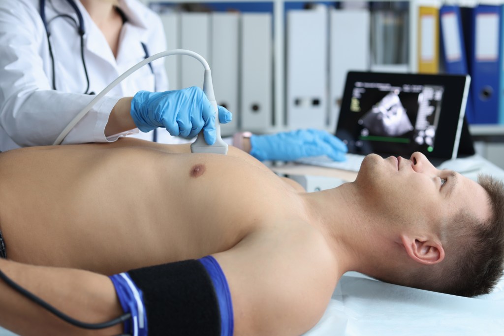
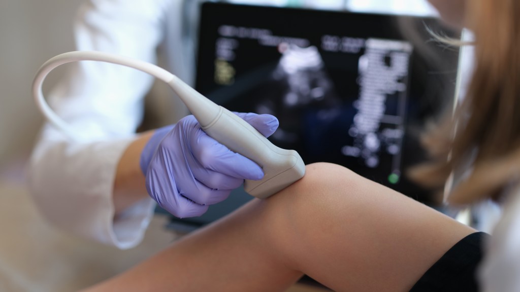
Ultrasound
Another commonly used imaging method is called ultrasound. It is a noninvasive test that sends sound waves into the body. Different types of tissue reflect sound waves differently. These differences can be picked up by an ultrasound machine and turned into a picture or video. Ultrasound is helpful for looking at the heart and other internal organs, other soft tissues such as blood vessels, or a developing baby.
Ultrasound enables healthcare providers to “see” details of soft tissues inside your body without making any incisions (cuts). And unlike X-rays, ultrasound doesn’t use radiation.
Although most people associate ultrasound with pregnancy, healthcare providers use ultrasound for many different situations and to look at several different parts of the inside of your body.
Other Scans
Doctors also use tests called nuclear imaging. These tests use a tiny amount of a radioactive substance, or “tracer.” Most tracers are injected into the body, but some are inhaled or swallowed. The tracers inside the body release radiation that can be measured by a detector outside the body. The type of tracer differs depending on what the doctors want to see.
A positron emission tomography (PET) scan, for example, often uses a radioactive sugar to diagnose cancer. When cancer cells take up the radioactive sugar, they can be seen with the PET scanner.
Scientists are working to develop new types of tracers to detect different conditions, such as infections hiding deep in the body. They’re also continuing to explore other ways to make medical scans faster and deliver less radiation.
A Note from QBan Health Care Services
Medical scans are very useful and generally safe imaging tests that healthcare providers use for a variety of reasons. If you need a medical scan and are worried about the exam or have questions about it, don’t be afraid to ask your healthcare provider. Talk to them about any concerns you have, including all options for testing and the benefits and risks of each. They’re available to help and support you.